Article Text

Statistics from Altmetric.com
Description
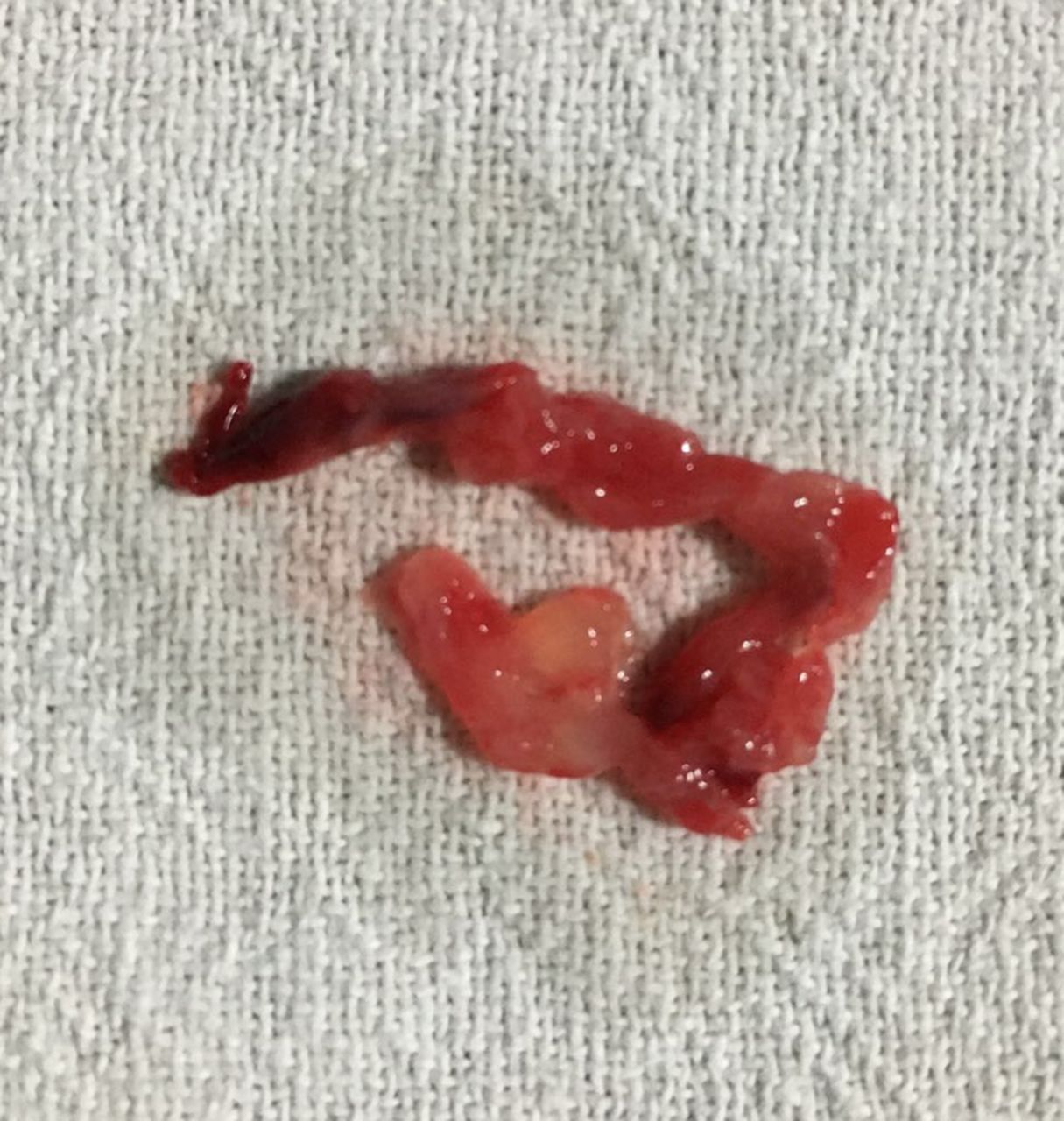
A 65-year-old man presented with acute-onset pain and numbness in the left upper limb for the past 6 hours. On examination, the left upper limb was pale and cold with absent pulses. There was no neurological deficit. The patient was a non-smoker, and had no atherosclerotic risk factors. Cardiac auscultation was normal. Subclavian artery angiography showed multiple filling defects in the proximal segment and total occlusion just after the origin of the vertebral artery (figure 1 and video 1). Transthoracic echocardiogram was carried out to assess a possible cardiac source for the embolism. A 0.9×1.2 cm rounded, heterogeneous, mobile mass was detected in the left atrium (figure 2 and video 2). The mass was attached to the interatrial septum in the region of the fossa ovalis. There was no associated disease of the mitral valve nor of the left ventricle. The location, mobility and echotexture suggested it to be a tumour rather than a thrombus. Urgent concurrent embolectomy of the left upper limb and open heart surgical removal of the cardiac mass were performed to avoid further embolism (figure 3). The diagnosis of atrial myxoma was confirmed by histopathological examination (figure 4). Myxomas constitute about 50% of all the benign cardiac tumours in adults and are mostly (>80%) found in the left atrium.1 Systemic embolisation occurs in around 30% of cases either from tumour fragmentation or complete tumour detachment. Cerebral embolism is the most common site followed by coronary, renal, mesenteric and peripheral arteries.2 Embolisation of cardiac myxoma to peripheral arteries leading to acute limb ischaemia has rarely been reported before.3 It should always be kept in the differential diagnosis of otherwise healthy patients who present with acute-onset limb ischaemia.
Learning points
Cardiac myxoma is the most common benign tumour of the heart and has high potential for systemic embolisation.
The most common site of embolisation is the central nervous system followed by the coronary, renal, mesenteric and peripheral arteries.
Embolisation to peripheral arteries causing acute limb ischaemia has rarely been reported before.
Cardiac myxoma should always be considered as a possible source of embolism in otherwise healthy patients presenting with acute limb ischaemia.
Management should aim at concurrent removal of both the embolus and cardiac tumour to avoid further embolism.

Subclavian artery injection showing a large filling defect in the proximal segment followed by total occlusion just after the origin of the vertebral artery.

Transthoracic echocardiogram, apical four-chamber view, showing the left atrial mass attached to the interatrial septum near the fossa ovalis. LV, left ventricle; RA, right atrium; RV, right ventricle.

Specimen removed during surgical embolectomy of left upper limb. It was soft and friable with a distinctive gelatinous appearance suggestive of a myxoma.

Histopathological examination showing myxoma with spindle cells with slender nuclei embedded in myxoid material with areas of haemorrhage and inflammation (H&E, ×40).
Transthoracic echocardiogram, Apical-4-chamber view, showing a highly mobile left atrial mass, which appears attached to the interatrial septum.
Subclavian artery angiography showing filling defects and total occlusion after the origin of the vertebral artery.
Footnotes
Competing interests None declared.
Patient consent Obtained.
Provenance and peer review Not commissioned; externally peer reviewed.