Article Text

Statistics from Altmetric.com
Description
A 55-year-old man was admitted to the emergency department, with high fever. Physical examination revealed a soft abdomen without any signs of peritoneal irritation and bowel sounds were normal. Laboratory tests revealed elevation of white cell count (11.5×10³/mm3) and C reactive protein. Contrast-enhanced CT showed sigmoid colon diverticulosis and increasing stranding of fat tissue adjacent to the sigmoid colon, in keeping with diverticulitis (figure 1). In addition, complete thrombosis of the inferior mesenteric vein (IMV) with intraluminal filling defect, perivenous inflammatory changes and subtle enhancement of the venous wall were identified (figure 2). There was no involvement of splenic and superior mesenteric veins, and intraluminal air was not present. A Gram stain of the patient's blood revealed the presence of Gram-negative bacilli. He was discharged and followed up on antibiotics and anticoagulation therapy.

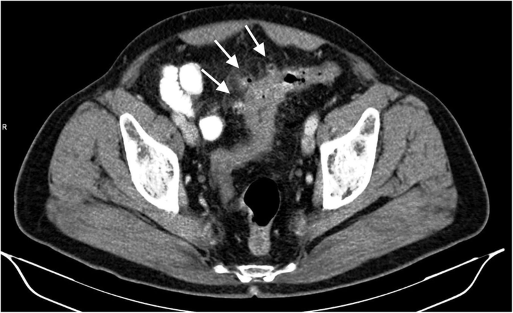
Contrast-enhanced axial CT image demonstrating multiple diverticula (arrows) and wall thickening of the sigmoid colon as well as surrounding fat stranding, in keeping with sigmoid diverticulitis.

Contrast-enhanced axial CT image showing pylephlebitis of the inferior mesenteric vein with hypodense filling defect through the lumen of the inferior mesenteric vein (arrows) and peripheral inflammatory changes (star).
Thrombophlebitis of the inferior mesenteric vein is commonly associated with the intra-abdominal inflammatory process and is also referred to as pylephlebitis.1 ,2 Sigmoid diverticulitis is the most common causative aetiology following other possible causes such as appendicitis, pelvic infections, pancreatitis and inflammatory bowel disease. The mortality rate is high in cases complicated by hepatic abscess or bowel ischaemia and infarction.3 Owing to its nonspecific signs and symptoms such as fever and nausea, imaging findings of IMV thrombophlebitis are very essential in its accurate diagnosis. CT is the diagnostic tool, with its high sensitivity in displaying intraluminal filling defects and underlying intra-abdominal inflammatory processes. Furthermore, life-threatening complications accompanied by IMV pylephlebitis can also be determined precisely by CT.
Learning points
Septic thrombophlebitis of the inferior mesenteric vein is a rare complication of the intra-abdominal inflammatory process and requires early diagnosis to prevent life-threatening complications. Radiologists must be aware of this fatal complication and assess the mesenteric venous system using contrast-enhanced CT.
Footnotes
Contributors CY reviewed the literature and wrote the manuscript. MA and MMO revised and edited the manuscript.
Competing interests None declared.
Patient consent Obtained.
Provenance and peer review Not commissioned; externally peer reviewed.