Article Text

Statistics from Altmetric.com
Description
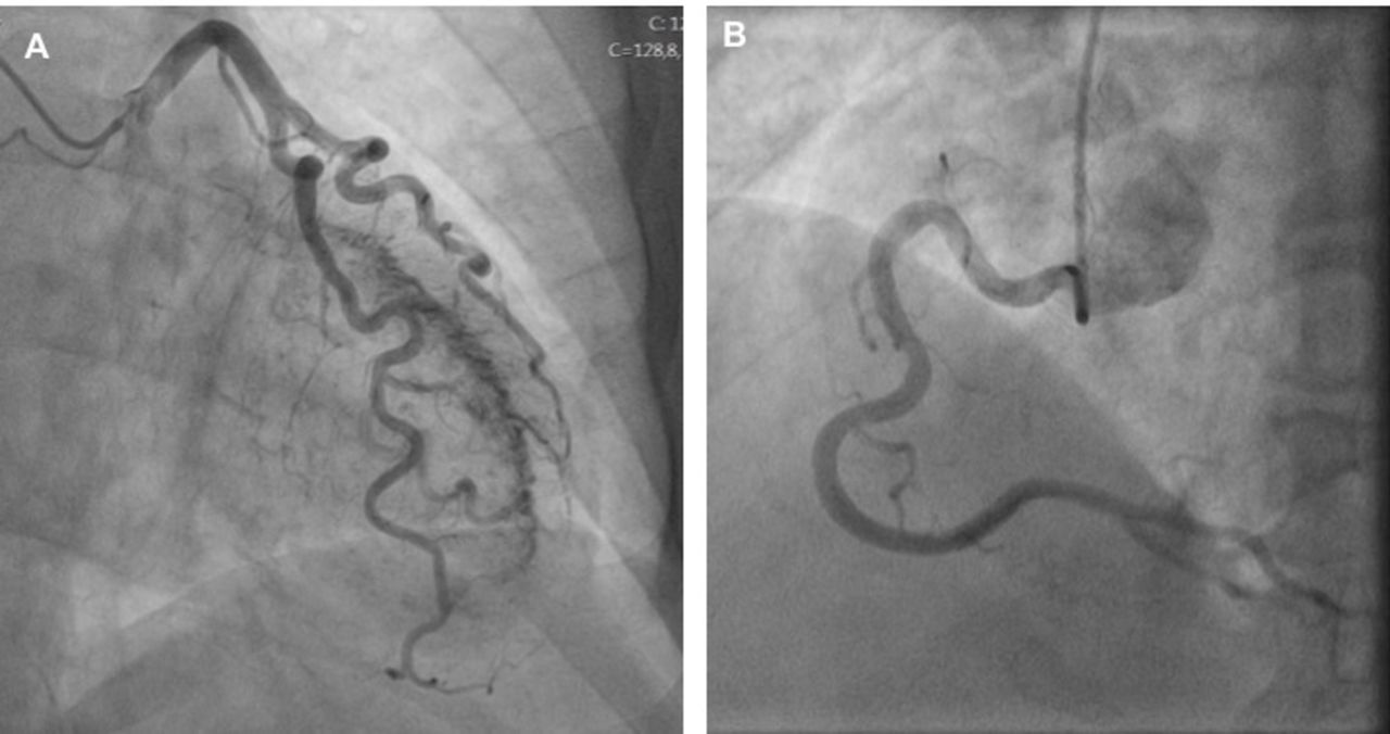
The authors present the case of a 74-year-old woman with a previous history of hypercholesterolaemia, hypertension and atypical chest discomfort, who was admitted to the emergency department with severe retrosternal pain radiating to her left shoulder and dyspnoea of 2 hours of onset. On admission, physical examination was normal, with no signs of cardiac failure. The ECG showed a sinus rhythm with no pathological Q wave, abnormal ST segment or T wave changes. Laboratory blood tests showed elevated cardiac biomarkers (13 ng/mL for troponin and 42 IU/L for creatine kinase myocardial band). Transthoracic echocardiography presented normal wall motion with a left ventricular (LV) ejection fraction of 59% and mild LV hypertrophy. Due to clinical suspicion of coronary pathology, the patient was submitted to invasive coronary angiogram, which showed multiple microfistulas from the left coronary artery draining into the LV, with no significant atherosclerotic stenosis (figure 1A,B,video 1). Considering the numerous fistulas and the clinical context, the symptoms were thought to be caused by a coronary steal phenomenon. The patient was discharged for outpatient investigation with a follow-up coronary CT angiogram, that confirmed this rare congenital condition and excluded other cardiac anomalies. The patient was treated conservatively and at the follow-up, 1 year after the arteriography, the patient was asymptomatic.
Coronary angiography in the late phase of contrast injection showing multiple microfistulas from the left coronary artery draining into the left ventricle with no significant atherosclerotic stenosis. (A) Left coronary artery in a left anterior oblique cranial projection. (B) Right coronary artery in a left anterior oblique projection.
Coronary artery fistula (CAF) is a rare abnormality found in 0.3%–0.8% of the adult population referred for coronary angiography, with symptomatic CAF in older patients being seldom described.1 CAF can be congenital or acquired, resulting from trauma, infection or iatrogenic injuries. CAFs are subdivided into two types: solitary fistulas and coronary ventricular multiple microfistulas, with solitary fistulas being more common. Sixty per cent of these fistulas arise from the right coronary artery, and 90% terminate in the right side of the heart.2 3 The coronary angiogram of this patient showed multiple microfistulas from the left coronary artery draining into the LV (figure 1A,B; video 1). Thus, multiple myocardial fistulas that drain into the LV, as in this case, are rare.4 Clinical presentation will generally depend on the haemodynamic significance of the anomaly and most commonly CAFs are asymptomatic and are found incidentally. However, this abnormal connection may be an important cause of acute coronary syndromes due to decrease in coronary blood flow (coronary steal phenomenon) when the shunt is important, particularly in patients without atherosclerotic changes.5 The optimal therapeutic strategy for CAF is not clear due to the rarity of this condition. The best management strategy for asymptomatic patients is monitoring, since the incidence of late complications is very low. Symptomatic patients with focal fistulas with large shunts may benefit from closure of the shunt as early as possible. It is unlikely that multiple fistulas can be treated surgically, and therapy with beta-blockers can be used as an alternative, as in the case described. A cardiac MRI stress perfusion imaging was performed after the introduction of anti-ischaemic therapy to this patient, excluding myocardial ischaemia.
The authors present this case because coronary artery-LV multiple microfistulas are very rare, particularly in patients with advanced age.
Learning points
Coronary artery fistula (CAF) is a rare anomaly found in 0.3%–0.8% of the adult population referred for coronary angiography, with symptomatic CAF in older patients being seldom described.
CAFs are subdivided into two types: solitary fistulas and coronary ventricular multiple microfistulas, with solitary fistulas being more common. Sixty per cent of these fistulas arise from the right coronary artery, and 90% terminate in the right side of the heart. Thus, multiple myocardial fistulas that drain into the left ventricle, as in this case, are rare.
CAF may be an important cause of acute coronary syndrome due to decrease in coronary blood flow (coronary steal phenomenon) when the shunt is important, particularly in patients without atherosclerotic changes.
Acknowledgments
The authors thank Dr Regina Barros Pereira for her extremely valuable support during the preparation of this manuscript.
Footnotes
Contributors IDC and JM were responsible for medical data acquisition. IDC wrote the manuscript. PA was the doctor responsible for the case orientation. JC, PA and JM revised the manuscript for important intellectual content. All authors gave the final approval of the version to be published.
Funding The authors have not declared a specific grant for this research from any funding agency in the public, commercial or not-for-profit sectors.
Competing interests None declared.
Patient consent for publication Obtained.
Provenance and peer review Not commissioned; externally peer reviewed.