Ischaemic colitis
BMJ 2016; 355 doi: https://doi.org/10.1136/bmj.i6600 (Published 22 December 2016) Cite this as: BMJ 2016;355:i6600
Chinese translation
该文章的中文翻译

- J M Trotter, specialist registrar in general surgery1,
- L Hunt, specialist registrar in diabetes and endocrinology2,
- M B Peter, consultant general surgeon1
- 1Department of Surgery, Scarborough General Hospital, Scarborough YO12 6QL, UK
- 2Department of Diabetes, Endocrinology and Metabolism, Sheffield Teaching Hospitals, Royal Hallamshire Hospital, Sheffield S10 2JF, UK
- Correspondence to: J M Trotter j.trotter{at}nhs.net
What you need to know
Ischaemic colitis is different from mesenteric ischaemia or “ischaemic bowel”
Ischaemic colitis is typically acute in onset and has a high mortality rate
Patients with suspected ischaemic colitis need urgent admission to a gastroenterological unit with specialist surgical services
Some patients with ischaemic colitis can be managed conservatively
Computed tomography is the investigation of choice for initial diagnosis of ischaemic colitis, using colonoscopy within 48 hours to give further prognostic information and to confirm diagnosis
The incidence of ischaemic colitis1 has risen from 6.1 cases/100 000 person-years in 1976-80 to 22.9/100 000 in 2005-09.2 Acute gastrointestinal medical and surgical teams will see a few patients with ischaemic colitis each month. Prevalence increases with age and comorbidity,2 which might lead to an increase in the incidence of ischaemic colitis as the population ages.3 A small proportion of patients will present with a more chronic form of ischaemic colitis.
This article provides practical advice to non-specialists regarding the diagnosis, management, and guideline recommendations for ischaemic colitis in the acute setting.
What is ischaemic colitis and what causes it?
Ischaemic colitis and mesenteric ischaemia are different disorders but are often confused: table 1⇓ highlights their differences. Ischaemic colitis occurs when there is an acute, transient compromise in blood flow, below that required for the metabolic needs of the colon. This leads to mucosal ulceration, inflammation, and haemorrhage. The duration and severity of hypoperfusion determines whether the colonic injury is predominantly ischaemic or as a consequence of reperfusion.4Figure 1⇓ shows the arterial supply of the colon and the most common sites for ischaemic colitis.
Differences between mesenteric ischaemia and ischaemic colitis

Fig 1 Arterial supply of the colon and the most common sites for ischaemic colitis. The colon receives blood from both the superior and inferior mesenteric arteries. However, there are weak points, or “watershed” areas, at the borders of the territory supplied by each of these arteries,5 such as the splenic flexure and the transverse portion of the colon. These watershed areas are most vulnerable to ischemia when blood flow decreases, as they have the fewest vascular collaterals
Ischaemic colitis often has a multifactorial origin, and patients with extensive comorbidities are at particular risk. Box 1 lists common causes of ischaemic colitis.
Box 1: Common causes of ischaemic colitis
Physiological
Systemic—Heart failure, systemic inflammatory response syndrome (SIRS), atherosclerosis
Embolic—Atrial fibrillation
Thrombotic—Concurrent malignancy and haematological disorders6
Iatrogenic
Pharmacological—Chemotherapy, sex hormones, interferon therapy, pseudoephedrine, cardiac glycosides, diuretics, statins, non-steroidal anti-inflammatory drugs (NSAIDS), immunosuppressive drugs, vasopressors6 7
Surgical—Abdominal aortic aneurysm repair8
Endoscopic—Colonoscopy and bowel preparation media for colonoscopy4 9 10 11
What are the symptoms and signs of ischaemic colitis?
Acute presenting symptoms are commonly diarrhoea, rectal bleeding, and colicky abdominal pain.12 Examination typically reveals a soft abdomen with tenderness and voluntary guarding over the affected segment of colon. The presence of peritonitis suggests full thickness ischaemia, perforation, or alternative diagnosis. The acute onset of the symptoms is a useful distinguishing factor between ischaemic colitis and inflammatory or infective colitis, where abdominal pain often has a more insidious onset.13 Symptoms of ischaemic colitis manifest in a matter of hours and, unlike infective or inflammatory colitis, continue to worsen with systemic instability.
Ischaemic colitis may result in systemic inflammatory response syndrome (SIRS) with associated observations of tachycardia, hypotension, tachypnoea, and occasionally raised temperature without an infective focus. Patients can present in a state of shock, leading on to multiorgan failure if not treated correctly.
Clinically, it is difficult to differentiate between patients with possible infective, inflammatory, or ischaemic colitis, and even with diagnostic tests it is not always clear. Generalists need to be equipped to recognise patients with symptoms of colitis who are deteriorating and refer them for specialist opinion.
How do you diagnose ischaemic colitis?
Investigate patients with possible ischaemic colitis urgently. Computed tomography is the diagnostic investigation of choice. Guidance from the American College of Gastroenterology4 recommends that computed tomography is performed within the first few hours of admission, with care led by a senior clinician from this point. Colonoscopic evaluation is recommended within 48 hours to visualise mucosa and confirm diagnosis.
There is no role for abdominal plain radiographs or ultrasonography in diagnosing ischaemic colitis, though these investigations often used in practice in the assessment of abdominal pain. They can give some information about ischaemic colitis, such as “thumbprinting” on x ray or mural thickening and blood flow on ultrasonography and Doppler ultrasound.14 15 16 17 However, the same, and more, information is provided in greater detail on computed tomography that is not user dependent and is usually more readily available out of hours than ultrasonography.
Laboratory tests
In the presence of rectal bleeding, perform clotting studies and a haemoglobin level. Inflammatory makers such as C reactive protein and neutrophil count are likely to be raised. Check renal function as patients are at risk of acute kidney injury because of the inflammatory response in ischaemic colitis.
Serum lactate levels may be raised as a result of systemic dysfunction and hypoperfusion. The role of lactate in this scenario is in monitoring progress during resuscitation. Raised serum lactate does not indicate gastrointestinal ischaemia, and a normal lactate level does not exclude full thickness ischaemia of the colon.18
Contrast enhanced computed tomography
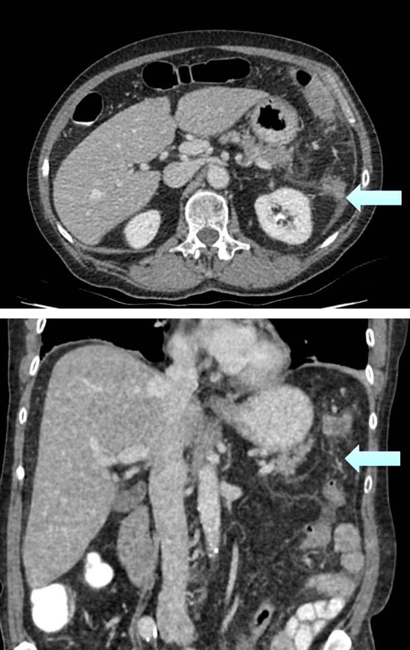
Computed tomography gives prompt information, with positive findings in ischaemic colitis in up to 98% of cases.19 These include wall thickening, abnormal or absent wall enhancement, dilatation, mesenteric stranding, venous engorgement, ascites, pneumatosis (gas within the bowel wall), and portal venous gas (fig 2⇓).19 20 The CT findings suggest a diagnosis of ischaemic colitis, but they can be present regardless of severity,19 limiting the prognostic value. The presence of such features (particularly in the watershed between the superior and inferior mesenteric artery) will suggest a diagnosis of ischaemic colitis but cannot absolutely distinguish it from other types of colitis. CT can rule out other diagnoses and complications such as perforation that will change management.

Fig 2 Computed tomographs of the abdomen (in axial and coronal views) showing fat stranding (increased density of fat, a sign of inflammatory process) and thickening (arrows) around the splenic flexure secondary to ischaemic colitis
Endoscopic evaluation
Early endoscopy can confirm the diagnosis by direct visualisation4 and provides prognostic information to help distinguish between cases that may settle with conservative management and those that may require emergency resection.
Transient non-gangrenous features of ischaemic colitis observed at colonoscopy include:
Petechial haemorrhages
Oedematous and fragile mucosa
Segmental erythema
Scattered erosions
Longitudinal ulcerations (colon single stripe sign) (fig 3⇓)
A sharply defined segment of involvement.21

Fig 3 Endoscopic findings of inflamed mucosa and single stripe sign (a single longitudinal strip of ulcerated or inflamed colon (arrow)) in segment of ischaemic colitis (reproduced with permission of www.natural-health-news.com)
Cyanosis and pseudo-polyps suggest a transmural ischaemia.
Colonoscopy is advocated by most studies, and there is no evidence that its use in assessment of ischaemic colitis is unsafe when performed by experienced practitioners.4 22 Retrospective studies of a total of 659 cases reported no cases of perforation secondary to colonoscopy,23 24 in data published in recent guidance.4
What treatment is available?
Initial resuscitation
There is no specific guidance for the resuscitation of patients with ischaemic colitis. General resuscitation principles apply, including
Intravenous fluid resuscitation
Fluid balance monitoring with bladder catheterisation
Assessment of acid-base status with arterial blood gas sampling
Blood glucose control and monitoring in diabetic patients.
While there is no specific evidence regarding fluid resuscitation in ischaemic colitis, aggressive and prompt resuscitation of a patient with SIRS has profound effects on outcomes, and specific algorithms now exist for conditions such as sepsis and pancreatitis.25 26
With appropriate resuscitation measures, colonic inflammation and associated symptoms settle in some patients without the need for surgery. Data on the proportion of patients who may be expected to settle without surgical intervention vary widely, reflecting the differences in clinical practice with regards to ischaemic colitis and the current lack of robust guidance.
Surgical intervention
Consider surgical intervention if there is radiological evidence of perforation, generalised peritonitis, or continuing haemorrhage causing instability or repeated transfusion. For patients without these features, decisions whether to operate when conservative management fails are made on an individual basis.
Factors associated with severe episodes that may not resolve with conservative treatment include4 22 27
Right sided distribution of colitis
Male sex
Lack of rectal bleeding
Renal dysfunction
Colonic strictures
Peritonitis.
Where one or more of these features exist, provide senior review daily and be alert to signs of development of full thickness ischaemia such as worsening pain or peritonism. For patients whose clinical condition is not improving, consider further blood texts to review biochemical markers. In the case of any clinical or biochemical deterioration, consider the need for repeat imaging and surgical intervention.
Patients who require surgical intervention for ischaemic colitis have higher mortality (37-48%4 17 22 27 28 29 30) than those treated conservatively (6.2% in a large systematic review17 22). Operative intervention usually includes segmental resection and colostomy formation. In unstable patients, complex surgery can worsen outcome.31
Caring for patients with ischaemic colitis
Anticoagulation
Prophylactic anticoagulation is advocated, but therapeutic anticoagulation is not indicated. Current guidance from the National Institute for Health and Care Excellence (NICE) advocates mechanical and pharmacological prophylaxis for venous thromboembolism for most groups of patients who don’t have contraindications, including those with ischaemic colitis.32 NICE guidance recommends postoperative prophylaxis for venous thromboembolism continues “until mobility is no longer significantly restricted.”
Cardiac emboli
Cardiac emboli have been found in 43% of patients with ischaemic colitis compared with 23% of matched controls.33 These findings may be coincidental, but consider investigations in those with cardiac symptoms or signs.33
Nutritional support
After admission for suspected ischaemic colitis, most patients will be fasted until a decision is made about surgery. There is a move away from prolonged fasting in modern surgical practice in acute and elective settings.25 34 35 Offer a dietetic-led enteral diet to help restore normal gut physiology and flora early. Parenteral nutrition may be necessary in severe cases when fasting is likely to exceed a week.
Antimicrobial therapy
The latest guidance on ischaemic colitis from the American College of Gastroenterology recommends antimicrobial therapy, although the evidence base for this is poor.4 Consider which specific agents to use with the help of microbiological guidance, taking account of local protocols and microbial resistance.
What is the long term management of ischaemic colitis?
Ischaemic colitis is multifactorial in origin and often occurs in a patient with multiple comorbidities. When treating ischaemic colitis, offer support in lifestyle modification to reduce recurrence or deterioration in other conditions, including advice on
Smoking cessation
Alcohol intake reduction
Increasing exercise.
There is no guidance or evidence to suggest that antiplatelets are of benefit in treating ischaemic colitis. It is not a purely atherosclerotic condition, so, alone, it is not a reason to start antiplatelet therapy.
Medication
Patients who have had ischaemic colitis may take regular medication that can impair colonic blood flow. These drugs are commonly prescribed for primary or secondary prevention of ischaemic heart disease such as angiotensin converting enzyme inhibitors or β adrenoreceptor blockers (see box 1). If cardiac medications have been stopped temporarily during the acute illness, reintroduce them with caution to avoid periods of hypotension that might exacerbate ischaemic colitis.
Follow-up care
Uncomplicated ischaemic colitis is usually followed up once after admission by the surgical team, then the patient discharged back to community care. Chronic or recurrent ischaemic colitis occurs in 6.8-16% of patients.4 This can present as another acute episode similar to the index admission. At the site of previous ischaemic colitis stricturing can occur, causing bloating, constipation, and colicky pain as well as chronic ulceration prone to bleeding that may manifest itself only as anaemia. The chronic, more benign symptoms of ischaemic colitis, though rare, are non-specific; if encountered, they warrant prompt referral to specialist services to confirm the diagnosis.
Methods
We searched the Medline database using the terms ischaemic colitis, ischaemia + colon, surgery + colitis. This yielded 1109 results that were reviewed for relevance and suitability. Most of the studies in this search were not primarily about ischaemic colitis and had no relevance to this review. Included studies were those of good methodology, acceptable analysis of data, and reasonable conclusions.
Questions for ongoing research
Does anticoagulation provide protection for recurrence of ischemic colitis?
Should Doppler ultrasound be more readily available in centres dealing with ischaemic colitis?
Should formal angiography and endovascular treatment be performed in mesenteric stenoses found on computed tomography of patients with ischaemic colitis?
Continuing medical education resources
These resources give further information and education on the subjects of colonic ischaemia and the recognition and management of patients, such as those with acute ischaemic colitis, who present with SIRS.
Colonic ischaemia
Brandt LJ, Feuerstadt P, Longstreth GF, Boley SJ; American College of Gastroenterology. ACG clinical guideline: epidemiology, risk factors, patterns of presentation, diagnosis, and management of colon ischemia (CI). Am J Gastroenterol 2015;110:18-44. www.nature.com/ajg/journal/v110/n1/abs/ajg2014395a.html
UpToDate. Colonic ischemia. www.uptodate.com/contents/colonic-ischemia?source=search_result&search=ischaemic+colitis&selectedTitle=1%7E56
Systemic inflammatory response syndrome
UpToDate. Sepsis syndromes in adults: epidemiology, definitions, clinical presentation, diagnosis, and prognosis. www.uptodate.com/contents/sepsis-and-the-systemic-inflammatory-response-syndrome-definitions-epidemiology-and-prognosis
Medscape. Systemic inflammatory response syndrome. http://emedicine.medscape.com/article/168943-overview
Information for patients
These resources provide information about ischaemic colitis that is written for people without formal medical training
Mayo Clinic. Ischemic colitis. www.mayoclinic.org/diseases-conditions/ischemic-colitis/basics/definition/con-20026677
Healthline. Ischemic colitis. www.healthline.com/health/ischemic-colitis#Overview1
How patients were involved in the creation of this article
No patients were directly involved in the creation of this article.
Footnotes
Contributors: JMT is lead author, LH is generalist advisor, and MBP is senior author.
Competing interests: We have read and understood BMJ policy on declaration of interests and have no relevant interests to declare.
Provenance and peer review: Not commissioned; externally peer reviewed.