Article Text

Abstract
Background Cornelia de Lange syndrome (CdLS) is a well known malformation syndrome for which five causative genes are known, accounting for ∼55–65% of cases. In this study, we hypothesised that mosaicism might explain some of the ∼35–45% of cases without detectable mutation in DNA derived from lymphocytes; we investigated the frequency of NIPBL mutations in buccal cells in individuals negative for mutations in any of the five genes in lymphocytes; and we evaluated the efficiency of obtaining DNA from buccal swabs and the best strategy for optimal mutation detection in CdLS.
Methods Buccal swabs were obtained from eight mutation positive and 13 mutation negative individuals with clinically diagnosed CdLS, following informed consent. We then forwarded instructions and a single mouth swab to the families; if subsequently insufficient DNA was obtained, we re-sent two mouth swabs. Buccal cells were screened for NIPBL mutations using Sanger sequencing techniques.
Results Sufficient DNA for analysis was obtained in 21/22 individuals. In all six tested individuals with a known NIPBL mutation and in two with a known SMC1A mutation, the mutation was confirmed in buccal cells. In 10 of the 13 tested individuals without detectable mutation in lymphocytes a NIPBL mutation could be detected in buccal cells. Clinically there were no significant differences between patients with a germline and mosaic NIPBL mutation.
Conclusions Somatic mosaicism for an NIPBL mutation is frequent (10/44; 23%) clinically in reliably diagnosed CdLS individuals. Obtaining buccal swabs at the time a blood sample is obtained will facilitate adequate molecular analysis of clinically diagnosed CdLS patients.
- Aetiology
- NIPBL
- SMC1A
- Buccal swabs
- genotype-phenotype
Statistics from Altmetric.com
Introduction
Cornelia de Lange syndrome (CdLS, or Brachmann-de Lange syndrome; OMIM 122470, 300590, and 610759) is a well known malformation syndrome characterised by a distinctive face, prenatal and postnatal growth retardation, limb malformations, and intellectual disability. To date, five causative genes have been identified: NIPBL, SMC1A, SMC3, RAD21, and HDAC8.1–6 Each one of these genes has a function in the sister chromatid cohesion process and CdLS is therefore termed as a cohesinopathy.7 Mutations in NIPBL are found in ∼50–60% of cases; the other genes account together for about 5% of clinically confirmed diagnoses, indicating that up until now CdLS can only be molecularly confirmed in ∼55–65% of patients (table 1).1 ,8–23 Studies by us and others using whole exome sequencing techniques failed to show pathogenic variants in CdLS individuals in whom mutations in the five known pathogenic CdLS genes had already been excluded (unpublished data). We hypothesised that this was caused by mosaicism and that searching for mutations using other tissues might yield additional mutations in genes known to cause CdLS.
Overview of studies describing results of mutation analysis in four or more individuals with clinically diagnosed CdLS*
We describe here the results of NIPBL mutation analysis in buccal cells in CdLS individuals without a detectable mutation in one of the five known genes in lymphocytes. Furthermore, we report on the efficiency of molecular analysis of buccal swabs, genotype–phenotype correlations in patients with and without mosaicisms, and discuss strategies for optimal mutation detection in CdLS.
Methods
Recruitment
In our earlier study9 we studied 39 CdLS individuals, to which we added five other CdLS individuals who were negative for NIPBL mutation analysis in lymphocytes. We asked eight mutation positive CdLS individuals described in the earlier study9 to participate by obtaining a buccal swab, to test for the reliability of molecular analysis of buccal swabs. All agreed. We then asked 17 individuals in whom no mutation was found in the five known genes to participate. Fourteen of them responded with consent.
We forwarded a single mouth swab to all families, asking parents to perform a buccal swab of their child. If insufficient DNA was obtained, we re-sent two mouth swabs to the families and asked them to repeat the procedure. No particular modifications were applied to increase the isolation of DNA from the swabs.
Severity scores
The severity score9 of the CdLS individuals in the earlier study was updated and the same severity score was added in the five patients who were not in the earlier study.
Molecular investigations
Genomic DNA was isolated from buccal swabs by using the Maxwell Buccal Swab LEV DNA Purification kit (Promega). Primers used for amplification of the 46 NIPBL coding exons (exons 2–47, NM_133433.3) and the corresponding exon–intron boundaries were designed using the Primer3 software (http://frodo.wi.mit.edu/primer3/). PCR fragments were sequenced using the Big Dye Terminator cycle sequencing kit v2 (Applied Biosystems), and analysed on a 3130 Genetic Analyser sequencing machine (Applied Biosystems). Sanger sequencing does not yield reliable quantitative results. The ratio between the variant and wild-type of a locus was evaluated by eyeballing only.
Ethics
The present study is part of a wider study in individuals with CdLS (‘CoDeLaGe’) and has been approved by the medical ethics committee of the Academic Medical Center in Amsterdam, and by the board of the Dutch CdLS support group.
Statistics
For analysis of correlations between ordinal categorical variables, the χ2 test for trend was used. Analysis was performed using SPSS V.20. The significance threshold was set at p<0.05.
Results
We obtained buccal swabs from a total of 22 individuals with CdLS and eventually sufficient DNA for mutation analysis could be harvested from 21/22. In five individuals we needed an extra pair of buccal swabs as the amount of DNA obtained from the first swab was insufficient. In one patient in whom we had found no mutation in lymphocytes, sufficient DNA could not be harvested from buccal cells despite collection of an extra set of buccal swabs.
In the total group of 44 individuals with CdLS we found 25 mutations in NIPBL, two in SMC1A, and none in the three other genes (SMC3, RAD21, HDAC8) (table 1). We were able to confirm in DNA derived from buccal cells the mutation found in NIPBL in all six individuals in whom such a mutation was earlier detected in DNA derived from lymphocytes (table 2); also the SMC1A mutation was retrieved in DNA isolated from buccal cells in the two tested CdLS individuals. Of 13 individuals with CdLS in whom no mutation was detectable earlier in lymphocytes, a mutation in NIPBL was found in buccal swabs in 10 of them (tables 2 and 3). The ratio between the pathogenic variant and wild-type was estimated to be about equal. As this mosaicism was unexpectedly high and might in theory point to an increase for NIPBL mutations in buccal cells irrespective of the presence of CdLS, we obtained a mouth swab from three healthy controls and excluded NIPBL mutations in them. Also the two CdLS individuals with an SMC1A mutation in lymphocytes were checked for an NIPBL mutation in buccal cells and were found to be negative.
Mutation detection rate in buccal swabs in relation to findings in lymphocytes
Mosaic NIPBL mutations detected in present CdLS cohort
We checked in DNA isolated from lymphocytes in each CdLS individual whether the variant detected in their buccal cells was present in the lymphocytes as well, by re-sequencing (Sanger sequencing) for that particular mutation, but none was retrieved (figure 1). In three CdLS individuals from the original group previously reported,9 no mutation was detected in either lymphocytes or buccal swabs.

Chromatogram showing the mutation c.358+3G>T in intron 4 identified in buccal DNA (lower lane) which is not present in lymphocyte DNA (upper lane).
The clinical characteristics of the CdLS individuals with a mutation detectable in lymphocytes, those with an NIPBL mutation detectable only in buccal swabs, and those without detectable mutation were compared using the severity score (table 4). The comparison is limited to the 37 CdLS individuals for whom we had sufficient data. Statistical analysis failed to show any significant difference between the group of individuals with a germline NIPBL mutation and mosaicism for an NIPBL mutation, and between the group of individuals with a germline NIPBL nonsense mutation and a mosaicism for an NIPBL nonsense mutation (p=0.704 and p=0.335, respectively). However, numbers were small and minor differences may have gone unrecognised.
Severity score features related to molecular findings
Discussion
Molecular confirmation of the clinical diagnosis of CdLS is of the utmost importance for adequate genetic counselling of families, and is critical in exploring genotype–phenotype correlations and for understanding the pathogenesis of the various manifestations of CdLS. We report here an unexpected and unusually high frequency of somatic mosaicism in CdLS individuals.
Mosaicism in CdLS has been reported before only infrequently: a chromosomal mosaicism was reported in 1965,24 and in 2010 a report of mosaicism for a c.2827delA mutation in NIPBL was published.25 The cohort of individuals with CdLS investigated in this study has been previously reported in an earlier genotype–phenotype study9 and a selection bias seems unlikely.
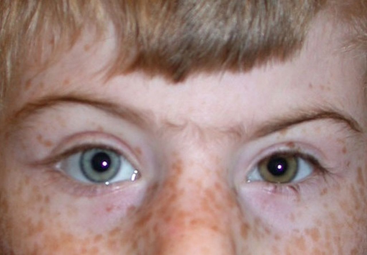
A similarly high frequency of mosaicism in a malformation syndrome with or without intellectual disability is unknown to us, except for entities that already show clear signs fitting mosaicism, such as asymmetries or pigmentation abnormalities.26–28 In the present series of people with CdLS a single individual showed a difference in colour between the left and right eye (figure 2), but otherwise none showed a significant clue for mosaicism. Heterochromia of the iris occurs in non-mosaic Mendelian conditions such as Waardenburg syndrome, but it is not a recognised sign in CdLS and must be very unusual as reports on many individuals with CdLS have been published. Heterochromia of the iris can occur in disorders caused by mosaic mutations such as Proteus syndrome, and therefore it seems possible that the heterochromia found in an individual mosaic for an NIPBL mutation is associated with the mosaicism. We cannot exclude, however, that its presence is coincidental.
Individual with Cornelia de Lange syndrome and a mosaic NIPBL mutation showing differently coloured irides.
There are several other malformation syndromes with intellectual disability, such as Rubinstein-Taybi syndrome and Kabuki syndrome, in which molecular confirmation of the clinical diagnosis is possible in only a limited percentage of cases, and we suggest performing similar studies in these entities. We have used only buccal swabs as second tissue to evaluate, purposes, but it has to be determined in each entity whether this is the right tissue to use. It might be that other easily available tissues such as bladder epithelial cells and hair bulbs are more suitable in other disorders. We do not exclude the possibility that further mosaicism can be detected in CdLS if other tissues are studied as well. Screening for mosaicism is especially important before initiating next generation sequencing studies (NGS) to detect additional pathogenic genes. If settings in evaluating whole exome sequencing studies are adequately set, one may be able to detect very low levels of mosaicism in NGS, but this would be an expensive approach.
The high rate of mosaicism for NIPBL mutations detected in the present study is remarkable and remains as yet unexplained. Theoretically the main mechanisms underlying this include somatic mutations (shortly) after fertilisation, loss of mutations in lymphocytes due to reversion, and selection against mutant cells specifically in lymphocytes.29 The absence of a difference in phenotype between CdLS individuals with a mosaic and germline NIPBL mutation argues against a somatic mutation after fertilisation. Reversion is a rarely detected phenomenon and mainly known with skin disorders, and would be unusually frequent for the various NIPBL mutations detected in the present study. We favour the hypothesis that there is a selection against lymphocytes with the mutation. This selection should take place specifically in lymphocytes and not in other easily available tissues. One may speculate an external influence such as acetylation of the cohesion complex to be of significance here.
Buccal swabs were shown to be an adequate way to obtain DNA from a second tissue in the present study. Swabs are cheap, can be performed at home by parents or other caregivers, and the success rates in obtaining sufficient DNA after one (14/20) or a repeat swab (5/6) or both (19/20; 95%) were high, despite the fact that NIPBL is a relatively large gene. The families did not consider taking one or two buccal swabs to be a significant burden.
The detection of a somatic mutation in a significant number of individuals (10/44; 23%) allowed us to detect a causative mutation in 37/44 individuals (84%), which is high compared to earlier reported studies (table 1). Possibly in these studies a significant number of cases had somatic mosaicisms as well. There is no significant difference in the classical CdLS signs and symptoms between individuals with a causative mutation detectable in lymphocytes and those with a mutation detectable in buccal cells (table 4), and it seems impossible to discern in advance those with and without somatic mosaicism. We restricted the present molecular analysis in buccal cells to sequencing of only NIPBL as it is by far the most frequently mutated gene in CdLS. We plan to perform further analysis in DNA derived from buccal cells for the other four genes known to cause CdLS as well. The first results of Sanger sequencing of SMC1A of DNA isolated from buccal cells of two CdLS individuals who were negative for the five known genes in lymphocytes and for NIPBL in buccal cells indicated no mutation was present. Further analysis is in progress.
An efficient and effective screening strategy to detect mutations in individuals with clinically diagnosed CdLS is important in daily patient care. We have adapted our diagnostic strategy and take a pair of buccal swabs in each CdLS individual together with the initial blood sampling. We sequence the buccal sample first for a NIPBL mutation; if negative we continue by sequencing the other four CdLS candidate genes in lymphocytes. If an NIPBL mutation is identified on buccal swab DNA, we then sequence NIPBL in DNA isolated from lymphocytes, because finding the mutation in both tissues will have consequences for the recurrence risk. We anticipate that with time NGS techniques will be used in diagnostics, using a targeted analysis of the results for variants in the five genes known to cause CdLS. Despite the high sensitivity of this technique to detect mosaicisms we have sincere doubts as to whether it will allow detection of mosaic NIPBL mutations in CdLS individuals. NGS of DNA isolated from buccal cells is in principle possible but technically demanding and unlikely to be available for patient care in the near future. Individuals who will be negative for both lymphocyte and buccal cell studies will be candidates for NGS using samples of both parents as well (trio strategy).
Conclusion
We conclude that there is a significant number of CdLS individuals who have somatic mosaicism for an NIPBL mutation. DNA derived from buccal cells using a buccal swab is a reliable way to investigate whether a patient may have a somatic mosaicism if lymphocyte analysis has failed to show a mutation. Obtaining a buccal swab at the time the initial blood sample is obtained will facilitate adequate molecular analysis of clinically diagnosed CdLS individuals.
Acknowledgments
The authors are grateful to the individuals with CdLS and their families for generously donating samples and clinical information. We thank all referring physicians and the Dutch CdLS Support Group for their cooperation, and the Prinsenstichting and Academic Medical Center for their support.
References
Footnotes
SAH and EJWR contributed equally.
-
Contributors Concept and design: RCMH. Gathering patient samples, clinical studies in patients: SH, SM, RCH. Molecular studies: EJWR, MMM. Drafting manuscript: SAH, EJWR, RCMH. Final approval: all authors.
-
Funding The study was supported by the Academic Medical Center and Prinsenstichting.
-
Competing interests None.
-
Patient consent Obtained.
-
Ethics approval Medical Ethical Committee, Academic Medical Center, Amsterdam.
-
Provenance and peer review Not commissioned; externally peer reviewed.
-
Data sharing statement All original data not mentioned in the manuscript will be made available to others upon request.