Article Text

Abstract
Background Flow-diverting stents have demonstrated great promise for the treatment of cerebral aneurysms; however, clinical experience with the devices remains very preliminary. We present two cases of spontaneous delayed complications—one fatal aneurysm rupture and one symptomatic increase in aneurysm volume—following the treatment of intradural aneurysms with the Pipeline Embolization Device (PED).
Presentation/intervention Two patients with unruptured, intradural aneurysms of the carotid artery underwent uneventful treatment with the PED (eV3, Irvine, California, USA). One patient, with a giant aneurysm of the carotid terminus, experienced worsening headache 5 days after the procedure and ultimately collapsed and became unresponsive. CT of the head demonstrated acute subarachnoid and intraventricular hemorrhage. The patient died the following day. A second patient with a large left posterior communicating artery aneurysm presented with progressive memory loss 3 months after PED reconstruction of the carotid artery. Although serial CT angiograms showed progressive thrombosis of the aneurysm to near-complete occlusion, MR of the brain demonstrated marked interval growth of the collective aneurysm–intra-aneurysmal thrombus mass with extensive edema throughout the adjacent left temporal lobe.
Conclusions Flow-diverting devices have demonstrated tremendous promise for the treatment of complex, unruptured cerebral aneurysms. However, experience with this novel approach to aneurysm treatment is preliminary and the consequences of its application within the cerebrovasculature remain incompletely defined. Mural destabilization resulting in delayed, spontaneous, aneurysm growth and/or rupture may occur in the days to weeks following the application of flow-diverting devices to treat previously unruptured intracranial aneurysms. A better understanding of the incidence and etiology of these complications is essential for this technology to be optimally applied.
- Aneurysm
- complication
- device
- hemorrhage
- pipeline embolization device
- spontaneous rupture
- stent
Statistics from Altmetric.com
Introduction
Flow diverters represent the newest addition to the group of endovascular devices available for the treatment of cerebral aneurysms. Initial experience with these devices has been largely favorable with very high rates of complete aneurysm occlusion, few or no documented cases of deterioration in angiographic appearance after treatment, relatively low rates of procedural complications and short procedural times.1 2 Moreover, these devices have expanded our ability to achieve the constructive treatment of some of the most daunting cerebral aneurysms, many of which had been inadequately addressed with predicate surgical or endovascular technologies—for example, very large, giant, fusiform, circumferential and multiply recurrent aneurysms.1 2
The recent CE mark approvals of the Pipeline Embolization Device (PED; eV3, Irvine, California, USA) and Silk stent (BALT Extrusion, Montmorency, France) have led to much more widespread application of flow-diverting devices in Europe. As the utilization of these devices has rapidly increased, experience is quickly accruing and additional complications related to the application of this unique technology are becoming evident.3
We present two illustrative cases which reflect potential complications related to the application of this relatively new, endoluminal approach to cerebral aneurysm treatment.
Case report
To date we have treated a total of five patients at our institution using the PED. No patients have been treated with other flow-diverting devices. One patient suffered a post-procedural, perforator territory (pontine) infarct following reconstruction of a basilar aneurysm with two overlapping devices. In the remaining four patients, there were no intraprocedural or immediate (24 h) postprocedural complications encountered. Two patients experienced significant delayed complications (presented below). All treatments were performed in the UK following CE mark of the device.
Patient 1 (Figure 1)
Presentation
This individual presented with an initial complaint of increasing headache superimposed upon the background of a longstanding left homonymous hemianopsia. CT imaging demonstrated a giant right-sided aneurysm and she was referred for further diagnostic evaluation and treatment. Subsequent CT angiography (CTA) and catheter-based digital subtraction angiography confirmed a giant (35×23×25 mm) right carotid terminus aneurysm incorporating the origins of the M1 segment of the middle cerebral artery and A1 segment of the anterior cerebral artery. Following extensive discussions with both neurovascular surgeons and endovascular neurointerventionists, the patient elected to undergo endovascular treatment using a flow-diverting stent system.
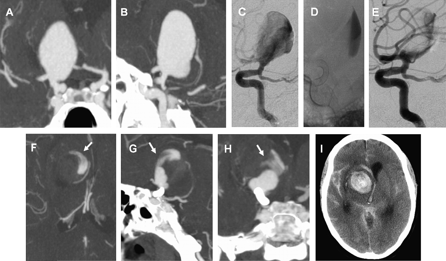
Patient 1: Coronal (A) and sagittal (B) reconstructions of CTA source data demonstrate a giant right internal carotid terminus aneurysm incorporating the origins of the M1 and A1 segments into the proximal fundus. Lateral subtracted angiogram (C) in the working angle for reconstruction demonstrates a well-organized flow jet directed toward the posterior dome of the aneurysm. Native image (D) after placement of a single Pipeline Embolization Device spanning from the proximal M1 segment of the middle cerebral artery proximally into the supraclinoid segment of the internal carotid artery. Following reconstruction, contrast layers dependently within the fundus of the aneurysm. Lateral subtracted angiogram (E) following treatment demonstrates a redirection of the primary inflow jet toward the more inferior aspect of the aneurysm fundus and reduced flow into the aneurysm overall. Five days after treatment the patient experienced severe headache. Repeat CT angiogram in the axial (F), sagittal (G) and coronal (H) planes demonstrated near-complete thrombosis of the aneurysm with residual filling along the anterior and medial aspect of the fundus. An irregular focus of contrast resembling a lamellated or cleft-like appearance extends into the more cephalad aspect of the thrombus mass (arrows), possibly indicative of acute hemorrhage penetrating into the immature thrombus mass. Later that evening the patient collapsed and became unresponsive. CT of the head performed after the ictus (I) demonstrates large volume subarachnoid and intraventricular hemorrhage as a sequela of aneurysm rupture.
Procedure
Dual antiplatelet medication was initiated with aspirin (75 mg once daily) and clopidogrel (75 mg once daily) administered for 1 week prior to treatment. Treatment was performed under general anesthesia through a right femoral access. A 6F Softip guiding catheter (Boston Scientific, Fremont, California, USA) was positioned within the distal cervical segment of the right internal carotid artery (ICA). Following placement of the guiding catheter, the patient was fully heparinized. The aneurysm neck was crossed with a Marksman catheter (eV3) and a Synchro-14 0.014-inch microwire (Boston Scientific) under high magnification fluoroscopic roadmap control. A 2.75×20 mm PED was then deployed uneventfully, spanning from the M1 segment distally, across the aneurysm neck and terminating within the supraclinoid segment of the ICA proximally. Immediately following placement of the PED, inflow into the aneurysm was clearly disrupted. Persistent dependent layering of contrast was observed (ie, an ‘eclipse sign’) at the end of the case. The PED appeared very well opposed to the parent artery and in an optimal position across the aneurysm neck. The patient emerged from general anesthesia neurologically intact and remained intact during the immediate postprocedural period. After the procedure, an intravenous heparin infusion was maintained for 1 day (25 000 U over 24 h) followed by 5 days of subcutaneous heparin (5000 U q12 h) at deep venous thrombosis prophylaxis doses. The patient was also maintained on dual antiplatelet medication.
Delayed complication
The patient remained well until the morning of the fifth postprocedural day when she complained of worsening headache. Repeat CTA demonstrated partial thrombosis of the aneurysm with persistent contrast filling of the medial aspect of the aneurysm fundus. In addition, contrast material extended in an irregularly crescentic, lamellated configuration from the residual patent lumen into the medial and cephalad aspect of the thrombus mass. Later that evening the patient lost consciousness and collapsed, requiring intubation and mechanical ventilation. A subsequent CT scan confirmed aneurysm rupture with high-volume subarachnoid and intraventricular hemorrhage. Surgical salvage was not attempted because of the patient's poor neurological condition. The patient never regained consciousness and died on the sixth postoperative day.
Patient 2 (Figure 2)
Presentation
This individual presented initially with staring spells noticed by their spouse. The patient was subsequently referred to a neurologist who initiated therapy with levetiracetam (Keppra; UCB Pharma, Inc., Smyrna, Georgia, USA) for presumed absence seizures and ordered brain MRI. The MRI demonstrated an 18×18×15 mm aneurysm arising from the posterior-lateral aspect of the distal left ICA with mass effect on the adjacent medial temporal lobe. Following review by a multidisciplinary neurovascular team the patient was offered vascular reconstruction with a flow-diverting stent.

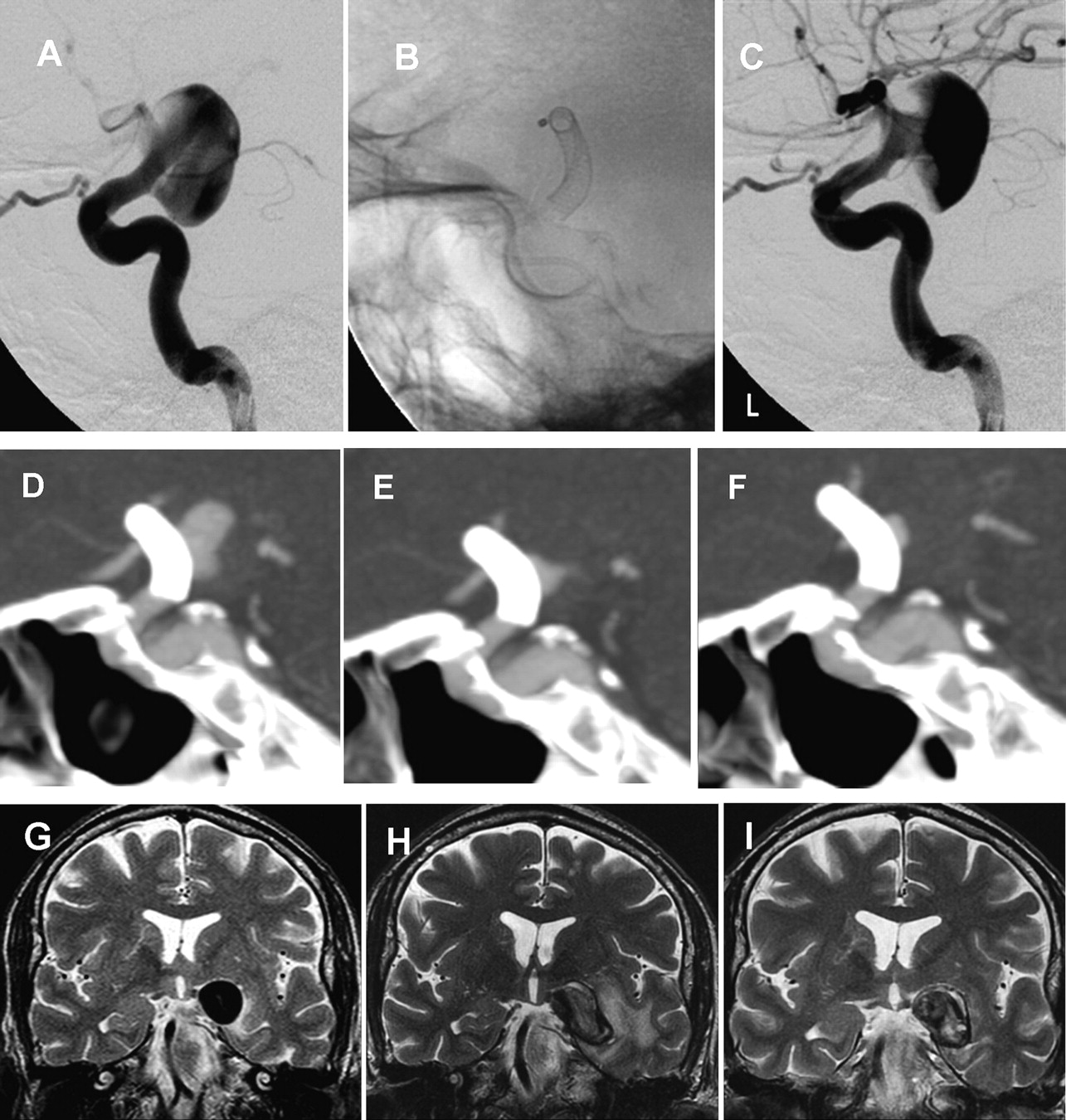
Patient 2: subtracted angiographic images performed in the working angles for treatment (A) demonstrate a very large posterior carotid wall aneurysm with a flow jet directed toward the superior posterior aspect of the fundus. Native image (B) following reconstruction shows a single Pipeline Embolization Device in place extending from the proximal M1 segment of the middle cerebral artery into the supraclinoid carotid artery, spanning the entire neck of the aneurysm. A subtracted image following treatment (C) shows disruption flow into the aneurysm with redirection of the primary inflow toward the more inferior aspect of the aneurysm fundus. Sagittal reconstructions of axial CT angiographic source data obtained 2 days (D), 1 month (E) and 3 months (F) after treatment demonstrate progressive thrombosis of the aneurysm with minimal filling in the region of the aneurysm neck and proximal fundus. Coronal T2 weighted sequence from an MRI examination before treatment (G) demonstrates a very large, posterior and laterally projecting aneurysm arising from the posterior wall of the supraclinoid internal carotid artery. The aneurysm exerts moderate mass effect upon the adjacent left temporal lobe. Three months after treatment, the patient noted a progressive deterioration in short-term memory. Coronal T2 weighted sequence (H) at a location analogous to the pretreatment MR shows interval enlargement of the aneurysm with progressive mass effect upon the left temporal lobe and extensive edema throughout the adjacent brain parenchyma. The initial images had shown a smoothly contoured lateral wall which at follow-up has taken on an irregularly, lobulated appearance. Clopidogrel was discontinued and the patient's symptoms improved. Follow-up MRI (I) shows near-complete resolution of the peri-aneurysmal edema with further evolution of the morphology of the lateral aspect of the aneurysm wall, which now appears less convex inferiorly. These progressive and continuing changes in the aneurysm wall morphology demonstrate the potential for the dynamic behavior of these partially thrombosed lesions after flow diversion.
Procedure
Dual antiplatelet medication was initiated with aspirin (75 mg once daily) and clopidogrel (75 mg once daily) administered for 1 week prior to treatment. Treatment was performed under general anesthesia through a right femoral access. A 6F Softip guider (Boston Scientific) was positioned within the distal cervical segment of the right ICA. Following placement of the guiding catheter, the patient was fully heparinized. The aneurysm neck was crossed with a Marksman catheter (eV3) and a Synchro-14 0.014-inch microwire (Boston Scientific) under high-magnification fluoroscopic roadmap control. A 3.5×16 mm PED was then deployed uneventfully, spanning from the M1 segment distally, across the aneurysm neck and terminating within the ICA proximally. The construct was judged to be in excellent position across the aneurysm neck with optimal wall apposition along the course of the parent artery. The patient emerged from general anesthesia neurologically intact and remained so during the immediate postprocedural period. After the procedure, an intravenous heparin infusion was maintained for 1 day (25 000 U over 24 h) followed by 5 days of subcutaneous heparin (5000 U q12 h) at deep venous thrombosis prophylaxis doses. The patient was also maintained on dual antiplatelet medication.
Delayed complication
The patient was discharged home on postoperative day 2. CTA prior to discharge showed substantial thrombosis of the aneurysm with a small residual remaining in the region of the neck. One-month follow-up CTA demonstrated a small volume only of persistent aneurysm filling the same location. Follow-up digital subtraction angiography was planned for 6 months post-treatment. Three months after treatment the patient began experiencing progressive worsening of short-term memory. While CTA demonstrated no apparent change in the degree of aneurysm filling, both the CTA and a subsequent MRI showed interval enlargement of the aneurysm–thrombus mass with worsening mass effect and extensive vasogenic edema throughout the left medial temporal lobe. The lateral margin of the aneurysm, which previously had a smoothly contoured ovoid appearance, had progressed to an irregular and lobulated configuration. The patient was instructed to immediately cease taking clopidogrel with the intention that the small volume of residual aneurysm filling might go on to complete thrombosis under these conditions. Repeat MRI 6 weeks and 3 months later demonstrated stabilization of the process with some resolution of the associated mass effect and temporal lobe edema. CTA showed persistent residual filling of the aneurysm along the medial aspect of the neck. The patient subjectively reports that his memory impairment has started to improve.
Discussion
Initial experience with flow-diversion technology has been largely positive. However, until very recently, these devices had been only used in a small and highly selected group of patients.1 2 4 Since the technology has now been approved for commercialization in the European Union it has been increasingly applied to treat a much larger population of patients with cerebral aneurysms. As this clinical experience accumulates, the efficacy of the flow-diversion treatment strategy as well as its consequences and limitations are starting to become more evident.
In the current report we discuss two cases which illustrate unexpected adverse effects that are likely related to the application of flow-diversion technology for the treatment of very large or giant intracranial aneurysms. In both cases, the flow-diversion treatment strategy not only failed to secure the aneurysm but also potentially contributed to a loss of the structural integrity of the aneurysm wall—leading to precipitous growth in one case and fatal rupture in the other.
While these represent two of a total of only five cases performed at our center, the actual incidence of this complication is not accurately reflected by these numbers since other institutions have reported much larger case series and have not observed these phenomena.4 5 There have been recent publications and conference papers reporting similar complications following flow diversion by other operators at different institutions. As such these are certainly not isolated or center-specific observations.6–9 Delayed rupture after flow diversion also does not appear to be specific to the particular ‘brand’ of flow diverter (eg, Silk or Pipeline). Given the lack of organized data collection within the context of commercialization thus far, the incidence of delayed aneurysm growth and rupture is impossible to estimate from the available information.
Similarly, the etiology by which destabilization of the aneurysm wall occurs following flow diversion remains completely speculative at this point. Important factors potentially include some combination of a redirection of the aneurysm inflow jet, an alteration of intra-aneurysmal shear forces to new points within the fundus and inflammatory and/or ischemic effects induced by the accumulating acute intra-aneurysmal thrombus upon the aneurysm wall. Both factors may have played a role in each of the present cases since the flow-diversion strategy induced partial thrombosis within the aneurysm fundus prior to subsequent growth/rupture and in both cases some intra-aneurysmal flow persisted after treatment. In both patients, a loss of the structural integrity of the aneurysm wall after treatment is demonstrated, by rupture in one case, and by marked growth and the development of a very irregular fundal contour with regional edema in the other.
Strikingly similar findings demonstrating the potential for a toxic effect of thrombus upon an aneurysm wall have been observed in the setting of partially thrombosed abdominal aortic aneurysms (AAAs). AAAs have been found to be at higher risk of growth and rupture when they are associated with a large volume of intraluminal thrombus.10–12 Histopathological studies of AAAs have shown that aneurysm wall segments that are covered by thick intraluminal thrombus are typically thinner, have fewer smooth muscle cells, fewer elastin fibers (which are more likely to be fragmented) and larger numbers of inflammatory cells when compared with wall segments that are covered by little or no thrombus.13 Intraluminal thrombus has also been demonstrated to induce local hypoxia of the aneurysm wall, which can lead to a loss of structural integrity.14 15 Interestingly, the CTA findings of irregularly lamellated flow penetrating into the intraluminal thrombus mass seen in patient 1 immediately prior to rupture (figure 1) closely resemble the ‘high attenuating crescent sign’ that has been associated with the impending rupture of an AAA on non-contrast abdominal CT.16 This observation may be directly analogous to the ‘active extravasation’ of contrast into an intraluminal thrombus mass that has been described in the same clinical setting with abdominal CTA.17
One issue that seems evident from these cases is that any residual flow within an aneurysm after attempted flow diversion preserves the potential for aneurysm growth and/or rupture. If evolving thrombus does, in fact, result in a destabilization of the aneurysm wall in some cases, partial occlusion of an aneurysm after flow diversion may create a situation that is unstable and potentially more dangerous than the completely patent aneurysm prior to treatment. Correspondingly small amounts of residual filling in unruptured aneurysms treated with flow diversion as monotherapy cannot necessarily be viewed in the same context as the small ‘neck remnants’ that are detected following incomplete clipping or endosaccular coiling of unruptured aneurysms—that is, aneurysms with small ‘neck remnants’ following conventional treatment are generally associated with a relatively low risk of rupture. Furthermore, patients treated with flow diverters are commenced on antithrombotic therapy, which may exacerbate the severity of any subsequent hemorrhage. Thus, in the absence of mechanical reinforcement of the aneurysm fundus, complete aneurysm occlusion seems to be a necessity (ie, the only ‘acceptable’ and safe endpoint) after flow-diversion monotherapy.
In both of the index cases, the aneurysm neck either partially or completely incorporated the carotid terminus. It is possible that persistent outflow demand into the jailed anterior cerebral artery contributed to the continued patency of the aneurysm neck, inhibiting progression to complete occlusion. These cases suggest that the flow-diversion strategy should not be applied to aneurysms that have incorporated branch vessels since continued flow through these branches may prevent aneurysm thrombosis, and perpetuate the adverse effects of the developing intra-aneurysmal thrombus upon the aneurysm wall. Given that the etiology of the observed mural destabilization is unclear, the impact of any proposed preventative measure—for example, steroids to reduce aneurysm wall inflammation during thrombosis—is not predictable. Theoretically, the most straightforward technical means by which to reduce, if not prevent, these problems may be to perform coil embolization of the aneurysm sac prior to, or using a parallel technique after, the placement of the flow-diverting construct. In approximately half of the aneurysms treated within the Pipeline for the Intracranial Treatment of Aneurysms (PITA) trial coils were in place within the aneurysm in addition to the endoluminal flow diverter, and no spontaneous delayed aneurysm rupture or growth was observed in any of the 31 patients enrolled.5
Finally, it is important to acknowledge that throughout the history of vascular neurosurgery, the treatment of very large and complex aneurysms has been fraught with hazards, some of which still remain poorly understood. Strategies such as Hunterian ligation as well as modifications of the technique described by Halsted18 and Neff,19 partial inflow occlusion20 and ‘high flow’ bypass have been applied in an attempt to achieve the treatment of otherwise ‘untreatable’ aneurysms.21 While there have been successes reported with each of these techniques, there have also been a considerable number of unforeseen failures resulting in parent artery thrombosis, stroke and delayed postoperative aneurysm instability, growth and rupture.22 As such, the present cases must be considered within the context of this very complex, poorly understood, unpredictable and highly lethal disease process.
Summary
Mural destabilization resulting in delayed, spontaneous, aneurysm growth and/or rupture may occur days to weeks after the application of flow-diverting devices to treat intracranial aneurysms. We hypothesize that the treatment of aneurysms with incorporated branch vessels may perpetuate intra-aneurysmal flow and predispose lesions toward delayed mural instability. The incidence and etiology of these complications must be better defined for this technology to be optimally applied.
References
Footnotes
Competing interests Dr Fiorella is an unpaid consultant and proctor for ev3/Chestnut Medical.
Patient consent Obtained.
Provenance and peer review Not commissioned; externally peer reviewed.