Article Text

Statistics from Altmetric.com
Description
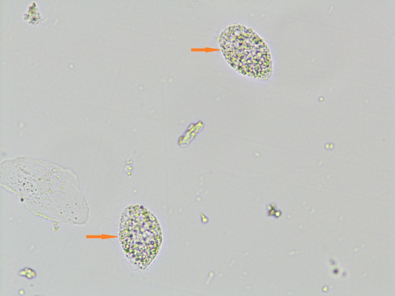
A 54-year-old female was admitted to the neurosurgery unit for excision of a non-functioning pituitary adenoma. Preoperative microscopic urinary examination revealed many motile pear- shaped organisms ranging in size from 17 to 23 µm (figure 1, red arrow) demonstrating characteristic wobbling to rotatory movement (video 1). These were conforming to the morphology of trophozoite form of Trichomonas vaginalis. Retrospectively, it was found that the patient had mild vaginal itching with passage of greenish discharge. Other laboratory investigations revealed that she was non-diabetic and HIV negative. Subsequently, she was given oral tinidazole 2 g single dose and was symptomatically better. She was taken up for surgery, and the follow-up urine sediment wet mount smears were negative for the parasite.
Trichomoniasis is the most common non-viral sexually transmitted infection worldwide. Vaginal/urethral/prostatic discharge and urine sediment wet mount smears offer rapid diagnosis with an excellent specificity owing to the characteristic morphology and motility of the organism.
Photomicrograph of urinary sediment smear showing two pear-shaped organisms (red arrows) conforming to the morphology of Trichomonas vaginalis trophozoites (wet mount, x600 magnification).
Numerous trophozoites of Trichomonas vaginalis demonstrating characteristic wobbling and rotatory motility in urinary sediment smears.
Learning points
Microscopic examination of the wet mount smears of the urinary sediment offers rapid diagnosis of active infection with Trichomonas vaginalis.
The characteristic morphology and the ‘wobbling’ and’ rotatory’ movement of the organism aid in specific diagnosis.
Footnotes
Contributors PS and PR: preparation of the manuscript. MUSS: correction and final approval of the manuscript.
Funding This research received no specific grant from any funding agency in the public, commercial or not-for-profit sectors.
Competing interests None declared.
Patient consent Obtained.
Provenance and peer review Not commissioned; externally peer reviewed.