Article Text

Statistics from Altmetric.com
Description
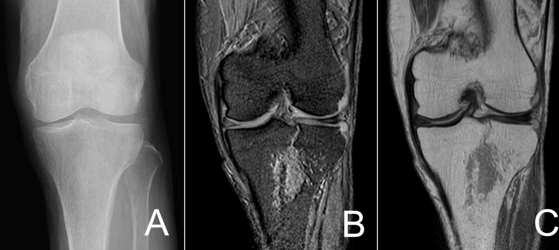
An 86-year-old woman presented to the emergency department with diffuse left knee pain from a fall from standing height. Anteroposterior and lateral X-ray of the left knee revealed no evidence of a fracture (figure 1A). However, MRI study revealed left tibial plateau fracture (coronal T2-weighted fast field echo imaging (figure 1B) and coronal proton density-weighted fast spin echo imaging (figure 1C). She was hospitalised for severe left knee pain and was given restrictions on weight-bearing activity for 2 weeks. She achieved good functional outcomes at a 6-month follow-up.
(A) An anteroposterior projection of left knee demonstrated no evidence of fracture. (B) MRI (coronal T2-weighted fast field echo imaging) revealed left tibial plateau fracture. (C) MRI (coronal proton density-weighted fast spin echo imaging) revealed left tibial plateau fracture.
Occult tibial plateau fractures are not be easily diagnosed on a radiograph. For occult tibial plateau fractures, non-operative conservative therapy should be a first-line treatment.1 However, these fractures can cause severe sequelae if they are not properly diagnosed and rapidly treated.2 When this occurs, patients may waste time and money compared with appropriate treatment. To prevent potential severe adverse events, MRI studies should be performed in patients with a suspected tibial plateau fracture.3
Learning points
Occult tibial plateau fractures may not be easily diagnosed on a radiograph.
MRI studies should be performed in patients with a suspected tibial plateau fracture.
Footnotes
Contributors The patient’s care was overseen by both the authors. YT provided assistance with the drafting of the manuscript. Both the authors approved the manuscript prior to submission.
Competing interests None declared.
Patient consent Obtained.
Provenance and peer review Not commissioned; externally peer reviewed.