Article Text

Statistics from Altmetric.com
Description
A 68-year-old man presented with cognitive impairment, urinary incontinence and short step gait. These symptoms, together with brain MRI finding, suggested idiopathic normal pressure hydrocephalus. After a spinal tap test to confirm the diagnosis, he suffered from postdural puncture headache. Despite an equivocal finding on spinal MR myelography (MRM), lumbosacral fat-suppressed T2-weighted image (FST2WI) clearly showed the characteristic ‘Dinosaur tail sign’ indicative of cerebrospinal fluid (CSF) leakage (figure 1).1 This pathognomonic sign disappeared on symptom improvement (figure 1).
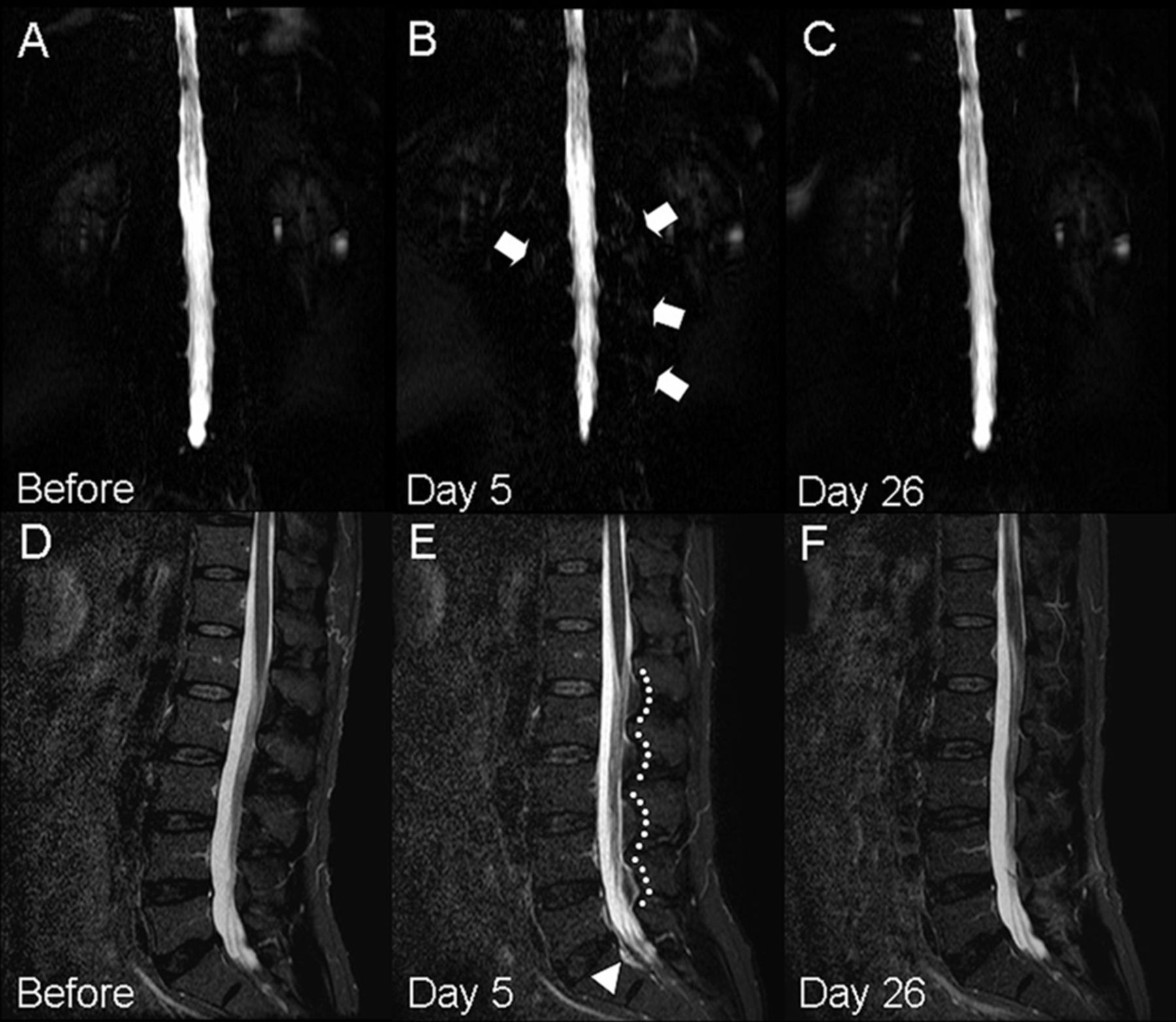
Serial MRM and FST2WI before (A, D) and after a dural puncture (B, C, E, F). In contrast to subtle paraspinal hyperintensities on MRM (arrows, B), sagittal FST2WI shows characteristic dorsal epidural fat tissues demarcated by interspinous arched hyperintensities (dash lines, E) contributing to the ‘Dinosaur tail sign’ and slight epidural hyperintensity (arrowhead, E). Pathognomonic signal changes disappear on symptom improvement (F). FST2WI, fat-suppressed T2-weighted image; MRM, MR myelography.
Spinal MRM is a radiation-free technique with excellent high-contrast resolution, making it possible to detect epidural fluid collections. Considering its non-invasiveness and high sensitivity for fluid signals, this technique should be used as the first-line examination in diagnosing CSF leakage.2 3 However, it could be hard to detect subtle epidural CSF leakage on spinal MRM using a two-dimensional sequence. On the other hand, sagittal FST2WI can be a useful technique to evaluate the subtle interspinous arched hyperintensities indicative of subtle epidural CSF leakage.
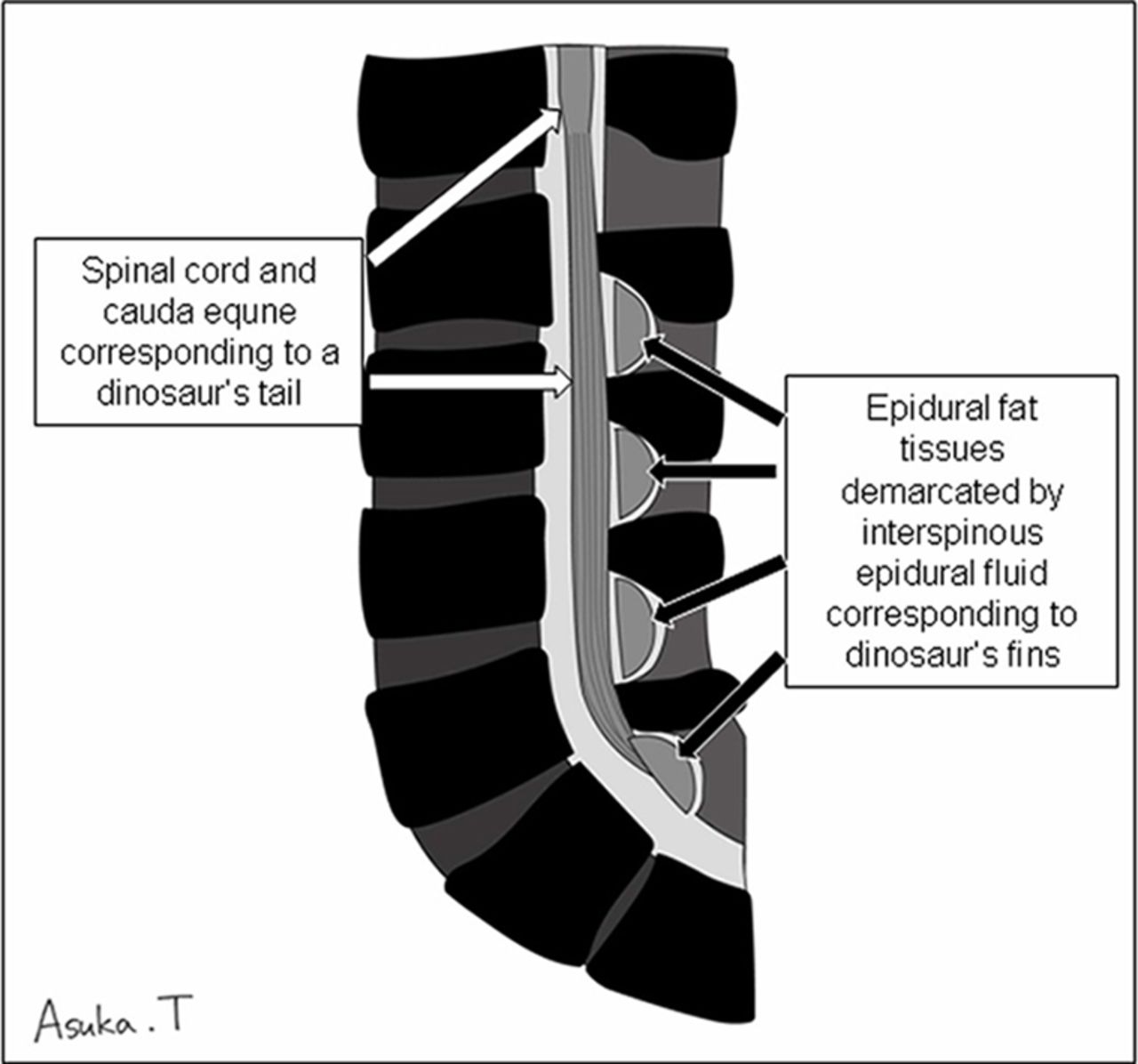
This case emphasises the use of ‘Dinosaur tail sign’ on lumbosacral sagittal FST2WI to reflect subtle extravasated epidural fluid collection to enable the accurate diagnosis of CSF leakage (figure 2).
A schema of the ‘Dinosaur tail sign’. This illustration represents that the combination of the spinal cord, cauda equine and dorsal epidural fat tissues resembles a dinosaur's tail and fins; therefore, this finding is termed as the ‘Dinosaur tail sign’.
Learning points
The ‘Dinosaur tail sign’ is a useful imaging finding indicative of subtle cerebrospinal fluid (CSF) leakage.
In addition to spinal magnetic resonance myelography, evaluation of subtle interspinous arched hyperintensities on lumbosacral fat-suppressed T2-weighted image is mandatory for the accurate diagnosis of CSF leakage.
Footnotes
Contributors KS: case report concept, interpretation of data and critical revision of manuscript for intellectual content. SM: interpretation of clinical data. MK: interpretation of imaging data. MM: acquisition of data, collection and assembly of data, and case report supervision.
Competing interests None declared.
Patient consent Obtained.
Provenance and peer review Not commissioned; externally peer reviewed.