Article Text

Statistics from Altmetric.com
Description
Talon cusp is a comparably rare developmental dental anomaly assumed to arise because of evagination on the surface of a tooth crown during tooth calcification stage. Talon cusp also called as Eagle's is a well-defined extra cusp like structure located on lingual or palatal surface of deciduous or permanent anterior teeth. Its occurrence may be either unilateral or bilateral, single or multiple teeth in males or females. Prevalence of talon's cusp is reported to be between 0.06% to 7.7%. Eagle's talon is most commonly seen in maxillary lateral incisor >maxillary central incisor> maxillary canine> mandibular incisor.1
Mandibular talon cusp is a rare entity to occur. Mandibular talon cusps are more prevalent in men than women. Occurrence in talon cusp is more common in permanent dentition. Most of the mandibular talon cusp shows unilateral presentation. Talon cusp associated with permanent central incisor and primary lateral incisor are the most common tooth type in mandibular arch. Left sided talon cusp are comparatively more common in mandibular arch.1 Mandibular talon cusp may occur in isolation or with other dental anomalies such as hypodontia, hyperdontia, impacted teeth, peg-shaped incisor, dens invaginatus, cleft lip, gemination or fusion.
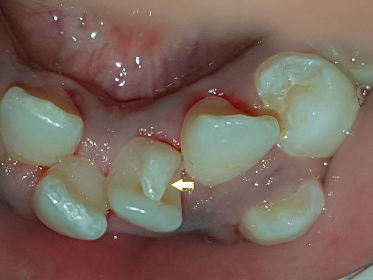
A 10-year-old boy presented to the clinic with a chief complaint of pain in lower back tooth for 7 days. Intraoral examination showed deep caries in right and left mandibular second deciduous molars. Further, a type I (Hattab classification 1996) talon's cusp was found in relation to mandibular right central incisor (figure 1).2 Intraoral periapical radiograph was used for confirmation of talon's cusp. (figure 2). Talon's cusp possesses both aesthetic and functional considerations. The management requires a sufficient knowledge of the present clinical entity and the problems associated with it. Management ranges from no treatment for small cusp, fissure sealant, composite restoration and prophylactic enameloplasty for deep grooves, to calcium hydroxide or Mineral Trioxide Aggregate pulpotomy or root canal therapy for severe cases.

Intraoral photograph showing Eagle's talon in right mandibular central incisor.

Intraoral periapical radiograph showing Eagle's talon in right mandibular central incisor.
Learning points
Early detection of talon's cusp is very important for the appropriate management.
Correct and preventive treatment plays a very vital role in avoiding the future complications.
Footnotes
Contributors PBK: case reporting, manuscript preparation, manuscript corrections, acquisition of data, interpretation of data, clinical diagnosis. RM: manuscript preparation and manuscript correction, clinical diagnosis. BD: interpretation of data, manuscript correction. PBJ: case reporting, manuscript preparation, acquisition of data.
Competing interests None declared.
Patient consent Obtained.
Provenance and peer review Not commissioned; externally peer reviewed.