Article Text

Statistics from Altmetric.com
Description
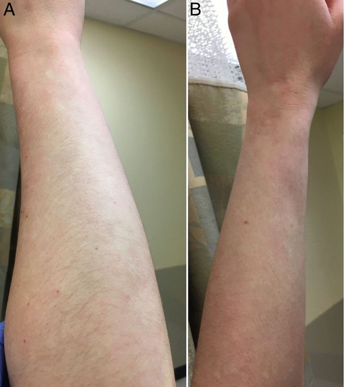
A 26-year-old woman with a reported history of tinea versicolour presented for persistent hypopigmentation on her bilateral forearms. Detailed examination revealed multiple small (5–10 mm), irregularly shaped white macules on the extensor surfaces of the bilateral forearms overlying slightly erythaematous skin. The surrounding erythaematous skin blanched with pressure and with elevation of the upper extremities the white macules were no longer visible (figures 1 and 2). A clinical diagnosis of Bier spots was made based on the patient's characteristic clinical features.

Patient's right forearm (A) and left forearm (B) showing multiple small symmetric hypopigmented macules against slightly erythaematous skin.
Hypopigmented macules fade and disappear when patient's right forearm (A) and left forearm (B) are raised.
Bier spots are completely asymptomatic and are often found on the extensor surfaces of the upper and lower extremities, although they are sometimes generalised.1 They are a benign physiological vascular anomaly, arising either from cutaneous vessels responding to venous hypertension or from small vessel vasoconstriction leading to tissue hypoxia.2 ,3 Our patient had neither personal nor family history of vascular disease.
Bier spots are easily diagnosed by a classic sign on physical examination: the pale macules disappear with pressure applied on the surrounding skin or by elevating the affected limbs (figure 2).1 However, Bier spots can be easily confused with a variety of other disorders associated with hypopigmented macules. The differential diagnosis includes vitiligo, postinflammatory hypopigmentation and tinea versicolour, which was a prior diagnosis in this case.1
Bier spots are often idiopathic and regress spontaneously, although there are reports of Bier spots heralding systemic diseases, such as scleroderma renal crisis, mixed cryoglobulinaemia or lymphoma.2 Since most Bier spots are idiopathic and transient, no treatment is required.1
Learning points
Bier spots are diagnosed clinically by the classic presentation of pale, irregularly shaped macules distributed on the extensor surfaces of upper or lower extremities, which disappear with pressure applied to or elevation of the affected extremity.
Bier spots can be easily confused with other dermatologic disorders with hypopigmented macules, including tinea versicolour.
As Bier spots are asymptomatic and often idiopathic, no treatment is required.
Footnotes
Contributors AH substantially contributed to the analysis and interpretation of data for the work, drafted the work, approved the final version and is accountable for all aspects of the work. SGK substantially contributed to the design and acquisition of data for the work, revised the work, approved the final version and is accountable for all aspects of the work. NK substantially contributed to the acquisition of data for the work, revised the work, approved the final version and is accountable for all aspects of the work. IB substantially contributed to the conception and acquisition of data for the work, revised the work, approved the final version and is accountable for all aspects of the work.
Competing interests None declared.
Patient consent Obtained.
Provenance and peer review Not commissioned; externally peer reviewed.