Article Text

Statistics from Altmetric.com
Description
A 94-year-old woman under our care receiving treatment for Clostridium difficile colitis reported abdominal pain and frequency of urination. She had a soft abdomen with mild suprapubic tenderness. She had a background of diverticulitis and a recent admission with C. difficile-related pseudomembranous colitis. An abdominal radiograph (figure 1) demonstrated a thin rim of air throughout the bladder wall. A CT scan (figure 2) further illustrated this finding, consistent with a diagnosis of emphysematous cystitis (EC). Urine microscopy showed mixed coliform species. EC is defined as air within the bladder wall and lumen, associated with infection.1 EC is a relatively rare condition that has a significant mortality rate of 7%.2 It has a diverse presentation ranging from an asymptomatic patient as an incidental finding on imaging to severe sepsis.2 A definitive diagnosis can only be made on abdominal imaging, and CT is recommended as the primary imaging modality if EC is suspected, to define the severity of the disease and exclude ascending infections.2 Risk factors include a neurogenic bladder, diabetes mellitus and being of female sex.2 It is important for physicians to be aware of how this condition appears on abdominal imaging in order to facilitate swift diagnosis and implement effective medical management including: broad spectrum antibiotics, tight glycaemic control, adequate bladder drainage and management of concurrent comorbidities, to avoid the morbidity and mortality associated with this condition.2 Our patient received a 5-day course of co-amoxiclav, and her symptoms and repeat abdominal radiograph demonstrated complete resolution of her EC.
Learning points
Emphysematous cystitis is an uncommon condition with a diverse presentation and a significant mortality rate of 7%.
Risk factors for this condition include diabetes mellitus, being of female sex and a neurogenic bladder.
Emphysematous cystitis can only be diagnosed definitively on abdominal imaging, and CT is recommended to assess severity and possible development of ascending infections.
Awareness of this condition is essential as medical management must be implemented early to prevent a poor outcome.
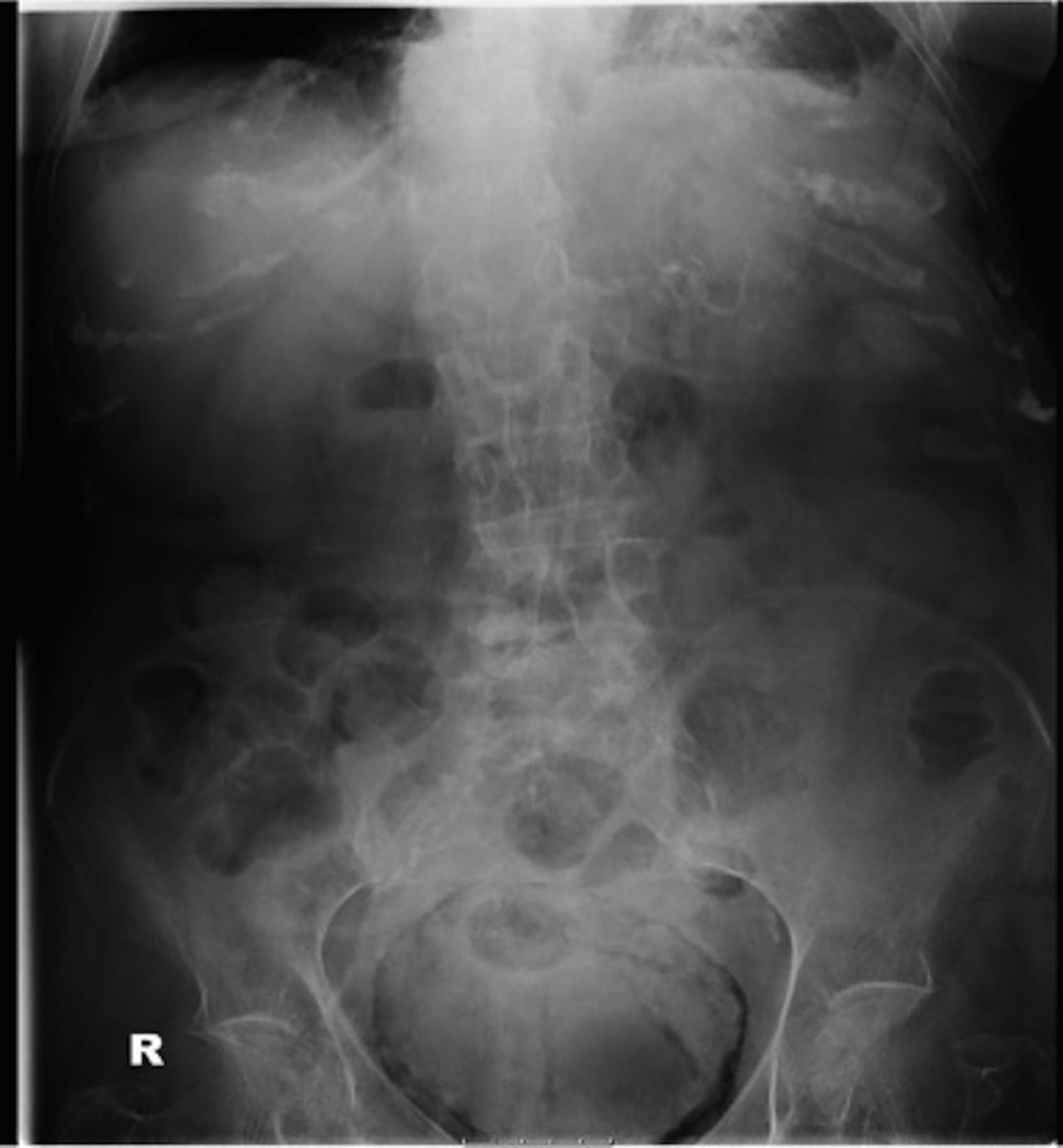
Abdominal X-ray demonstrating thin rim of air within the bladder wall.

CT image highlighting air throughout the bladder wall.
Footnotes
Contributors MI and ASB were involved in substantial contributions to the conception or design of the work, or the acquisition, analysis or interpretation of data. MI and OI were involved in drafting the work or revising it critically for important intellectual content. MI and OI were involved in final approval of the version published. MI, OI and ASB are in agreement to be accountable for all aspects of the work in ensuring that questions related to the accuracy or integrity of any part of the work are appropriately investigated and resolved.
Competing interests None declared.
Patient consent Obtained.
Provenance and peer review Not commissioned; externally peer reviewed.