Article Text

Statistics from Altmetric.com
Description
A 4-year-old girl presented with a 4-month history of progressive exotropia. There was no headache, no impaired upgaze, no nystagmus and no vision changes. Complete neurological examination was otherwise normal. MRI of the brain revealed a 1.5 cm cystic lesion in the pineal region. The cyst had a smooth margin lined by a thin wall of <1 mm that was homogenously enhancing on contrast scan. Compared to cerebrospinal fluid, the cyst content was isointense in T1 and hyperintense on T2-weighted images (figure 1). The pineal gland was slightly displaced anteriorly and laterally to the left without hydrocephalus. Given the unusual appearance with an associated new neurological deficit, an endoscopic biopsy and resection of the cyst was performed. Pathology demonstrated benign choroid plexus and neuroglial tissue without evidence of tumour (figure 2).

Sagittal T1-weighted image with contrast revealling a cystic lesion in the pineal region, with a thin wall that is homogeneously enhancing (A). On axial T2-weighted image, the cyst content is more hyperdense than cerebrospinal fluid and the pineal gland is displaced to the left (B) without evidence of reduced diffusivity on diffusion-weighted imaging sequences (C).
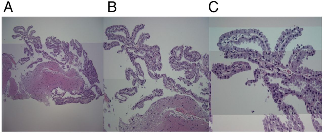
(A–C) Permanent histological sections obtained following biopsy revealling neuroglial tissue surrounded by benign choroid plexus projecting to the surface without evidence of neoplasm.
Pineal cysts are common incidental findings on brain MRI of children. Normally, the pineal cyst wall consists of an inner layer of glial tissue rich in Rosenthal fibres, residue pineal parenchyma and an outer fibrous layer.1 ,2 The presence of choroid plexus is very rare. Embryologically, the pineal gland arises from a diverticulum from the roof of the third ventricle. The distal tip of the diverticulum becomes a small cavity in the pineal gland, which may enlarge and become a cyst. One animal histology has shown choroidal-like cells in association with the cerebrospinal fluid (CSF)-contacting pinealocytes.3 Given the role of CSF production in the choroidal plexus, this close association may propose a mechanism of fluid production in the pineal cyst, which may mimic cystic pineal tumours.
Learning points
Pineal cyst is a common incidental finding in brain MRI of children. Surgery is generally indicated only when the patient is symptomatic or the lesion atypical.
Pineal cyst walls may comprise of an inner layer of neural glial tissue, residue pineal parenchyma and an outer fibrous layer, which may mimic tumours.
Footnotes
Twitter Follow Yuanfan Yang at @yangyuanfan@msn.com
Competing interests None declared.
Patient consent Obtained.
Provenance and peer review Not commissioned; externally peer reviewed.