Article Text

Statistics from Altmetric.com
Description
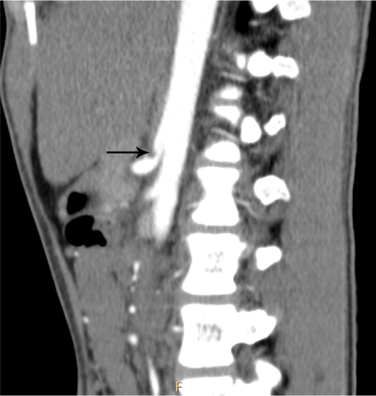
An 18-year-old man presented with a 7-month history of abdominal pain, particularly in the epigastrium, and weight loss of 14 pounds. Symptoms were aggravated post meals. An abdominal sonogram, upper gastroendoscopy and colonoscopy, were inconclusive. A contrast-enhanced CT of the abdomen revealed kinking of the proximal coeliac artery due to the median arcuate ligament, creating a hooked appearance with post-stenotic dilation (figure 1). A repeat sonography with Doppler revealed a normal calibre coeliac artery in inspiration with a peak systolic velocity (PSV) of 147 cm/s (figure 2A). On the contrary, there was colour aliasing suggestive of post-stenotic turbulence in expiration with a PSV of 304 cm/s (figure 2B). A diagnosis of median arcuate ligament syndrome was made. Surgical resection of the ligament led to relief of symptoms.

Contrast-enhanced CT of the abdomen, parasagittal reconstruction, showing kinking of the proximal coeliac artery, creating a hooked appearance (black arrow). There is post-stenotic dilation.

(A) Colour Doppler with spectral analysis of the coeliac artery in inspiration reveals normal calibre and a peak systolic velocity of 147 cm/s. (B) Colour Doppler and spectral analysis of coeliac artery on exhalation depicts colour aliasing (white arrow) suggestive of post-stenotic turbulent flow with a peak systolic velocity of 304 cm/s. There is widening of the spectral window.
The median arcuate ligament is a fibrous sling that courses superior to the coeliac trunk origin.1 When it is abnormally low, it compresses the artery, but symptoms of mesenteric ischaemia are rarely seen. A typical patient is a young female with postprandial pain and weight loss.1 CT reveals acute angulation and proximal narrowing of the coeliac trunk with post-stenotic dilation. This hooked appearance differentiates it from atherosclerotic narrowing.1 ,2 The narrowing accentuates on expiration, as evidenced by increased flow velocity and turbulence on Doppler.2 A PSV of over 200 cm/s during expiration or a ratio more than 3:1 of PSV of coeliac trunk to aorta in expiration is a Doppler criterion for diagnosis.2 The definitive diagnosis is, however, digital subtraction angiography.1 ,2
Learning points
Our case stresses the need to perform Doppler interrogation especially when grey scale findings are apparently normal. Screening of vascular causes of ischaemic pain can be easily performed with colour Doppler examination.
Classic angiographic findings of median arcuate ligament syndrome include kinking of the proximal coeliac trunk with post-stenotic dilation. The narrowing worsens when the patient exhales. Doppler study reveals increase in peak systolic velocity with turbulent flow on exhalation as opposed to inspiratory flow patterns.
Footnotes
Competing interests None declared.
Patient consent Obtained.
Provenance and peer review Not commissioned; externally peer reviewed.