Article Text

Statistics from Altmetric.com
Description
A 57-year-old female patient presented to our emergency department with irregular palpitations for more than 48 h. She had a history of hypothyroidism on treatment without any other cardiovascular risk factors. An ECG performed at admission revealed atrial fibrillation with rapid ventricular response. She was referred to our echocardiography laboratory for study. The transthoracic echocardiogram demonstrated a structurally normal heart. With a goal of electrical cardioversion, the patient underwent transesophageal echocardiogram (TEE), which revealed a 4 mm image of medium echogenicity, coating the entire left atrium and left atrial appendage (figure 1A–C).
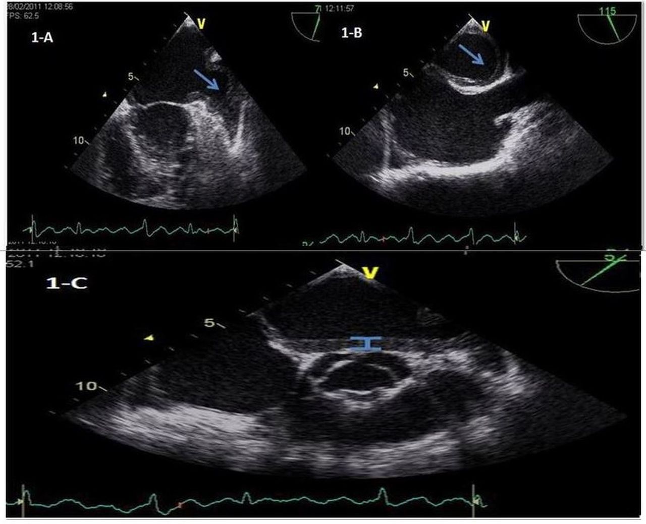
(A–C) Image of medium echogenicity coating the entire left atrium and left atrial appendage (arrows).
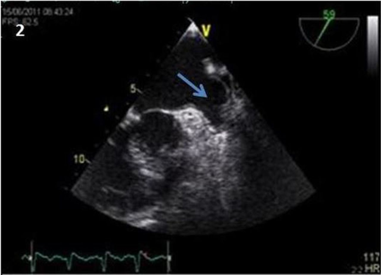
Considering the clinical context, a rather atypical left atrial thrombus was considered the most likely diagnosis and the patient was put on oral anticoagulation with vitamin K antagonist. Two months later, a novel TEE was performed and the thrombus had partially disappeared (figure 2). Thrombophilia was subsequently excluded.

Control transesophageal echocardiogram revealing the thrombus disappearance.
Thrombus formation in patients with non-valvular AF results from stagnant blood in the atria,1 combined with other physiological derangements such as progressive atrial dilation and abnormal platelet activation and changes in coagulation factors, contributing to an increased propensity for blood clot formation. This particular shape of thrombus coating the entire left atrium and left atrial appendage has never been reported before, and thus represents an extremely rare form of presentation. Physicians should be aware of the potential high variability of thrombus shape and location.
Learning points
-
Thrombus formation in patients with non-valvular atrial fibrillation results from stagnant blood in the atria, combined with other physiological derangements such as progressive atrial dilation and abnormal platelet activation and changes in coagulation factors.
-
Physicians should be aware of the potential high variability of thrombus shape and location.
-
Treatment with oral anticoagulant is extremely important in order to prevent embolisation.
Acknowledgments
The authors would like to thank Dr Providencia and Sergio Barra for help in the preparation of the manuscript.
Reference
Footnotes
-
Contributors AF was involved in writing the manuscript. JP reviewed the manuscript. JT and GC performed the ECG of the patient.
-
Competing interests None.
-
Patient consent Obtained.
-
Provenance and peer review Not commissioned; externally peer reviewed.