Article Text

Statistics from Altmetric.com
Description
An 87-year-old man was referred to our rheumatology clinic for assessment of painful, swollen and deformed fingers. He had a history of longstanding, but suboptimally treated gout, which had resulted in previous amputation of his left middle finger. On examination, there was marked digital deformity. The right index finger was erythematous, with multiple tophaceous deposits (figure 1). There was a severe loss of digital flexion and extension. Radiographs of both hands demonstrated punched-out juxta-articular erosions involving the metacarpophalangeal joints bilaterally (figure 2). There was marked soft tissue swelling with virtually complete osteolysis of the middle and distal phalanges of the index and little fingers of both hands. The right index finger was the most severely affected. Serum inflammatory markers and uric acid levels were within normal limits. On polarised microscopy of a right index finger aspirate, monosodium urate crystals were observed. A diagnosis of advanced erosive tophaceous gout was made. He was treated with long-term colchichine 0.5 mg twice per day and allopurinol 300 mg twice per day. A referral was made to the hospital's hand therapy service for functional optimisation. However, the patient's participation with rehabilitation was limited by medical comorbidities.
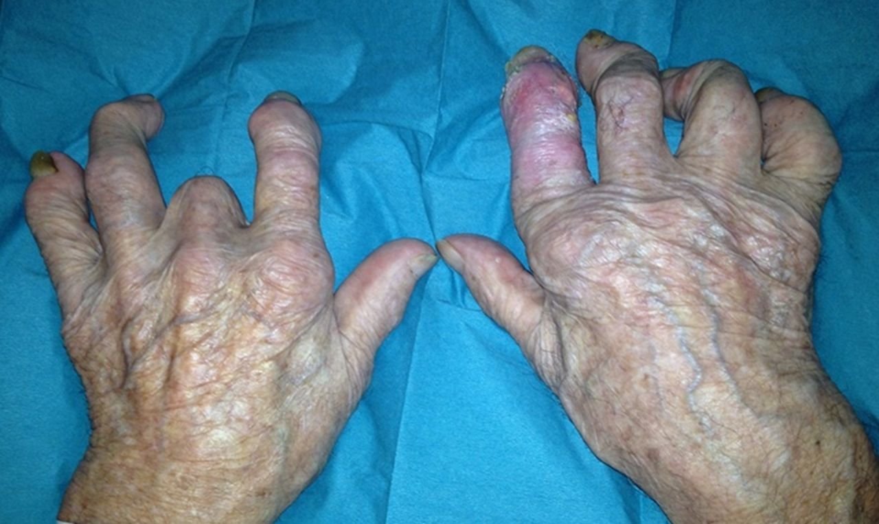
Clinical photograph of the patient's hands demonstrating a deforming polyarthropathy. There is marked erythema of the right index finger with yellow subcutaneous tophi visible and a previous amputation of the left middle finger.

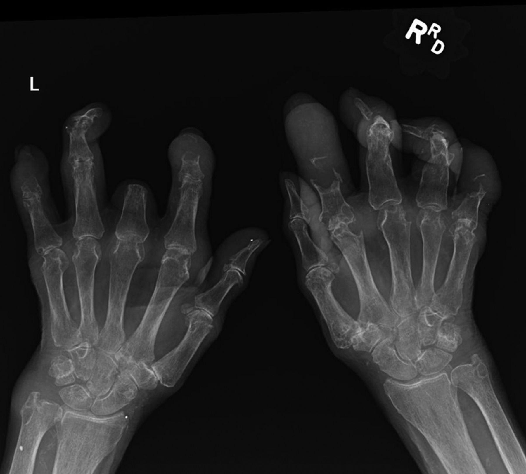
An anteroposterior radiograph of both hands demonstrating virtually complete osteolysis of the middle and distal phalanges of the index and little fingers of both hands. The skeletal architecture has been replaced with monosodium urate.
Radiographic features of advanced gout include tophaceous enlargement and ‘punched out’ erosions of juxta-articular bone.1 On review of the literature, we identified few cases of gross osteolysis,2 ,3 none of which demonstrated such extensive bone destruction as is seen in this case.
Learning points
-
Longstanding and untreated recurrent attacks of acute gout may lead to advanced tophaceous gout.
-
Severe tophaceous polyarticular gout can occur in the setting of a normal serum urate and inflammatory markers, and if doubt exists, a joint aspiration should be examined by polarised microscopy.
-
Monosodium urate deposition can lead to severe bony destruction and require digital amputation in rare cases.
Footnotes
-
Contributors CER, AB, PLR and KPO were involved in conception and design of the article. CER and AB contributed to the acquisition, interpretation of data and drafting of the article. PLR and KPO were involved in revising the article critically for important intellectual content and final approval of the version submitted.
-
Competing interests None.
-
Patient consent Obtained.
-
Provenance and peer review Not commissioned; externally peer reviewed.