Article Text

Statistics from Altmetric.com
Description
A 54-year-old man presented with claudication in his left calf with a walking distance of 200 m. There was no history of hypertension, hyperlipidaemia, thrombophilia, smoking or any risk factors associated with atherosclerosis.
Doppler scan demonstrated occlusion from the distal superficial femoral artery (SFA) to the proximal popliteal artery. A lower limb MR angiography supported this and showed significant collateralisation (figure 1). He underwent CT angioplasty with immediate symptomatic relief.
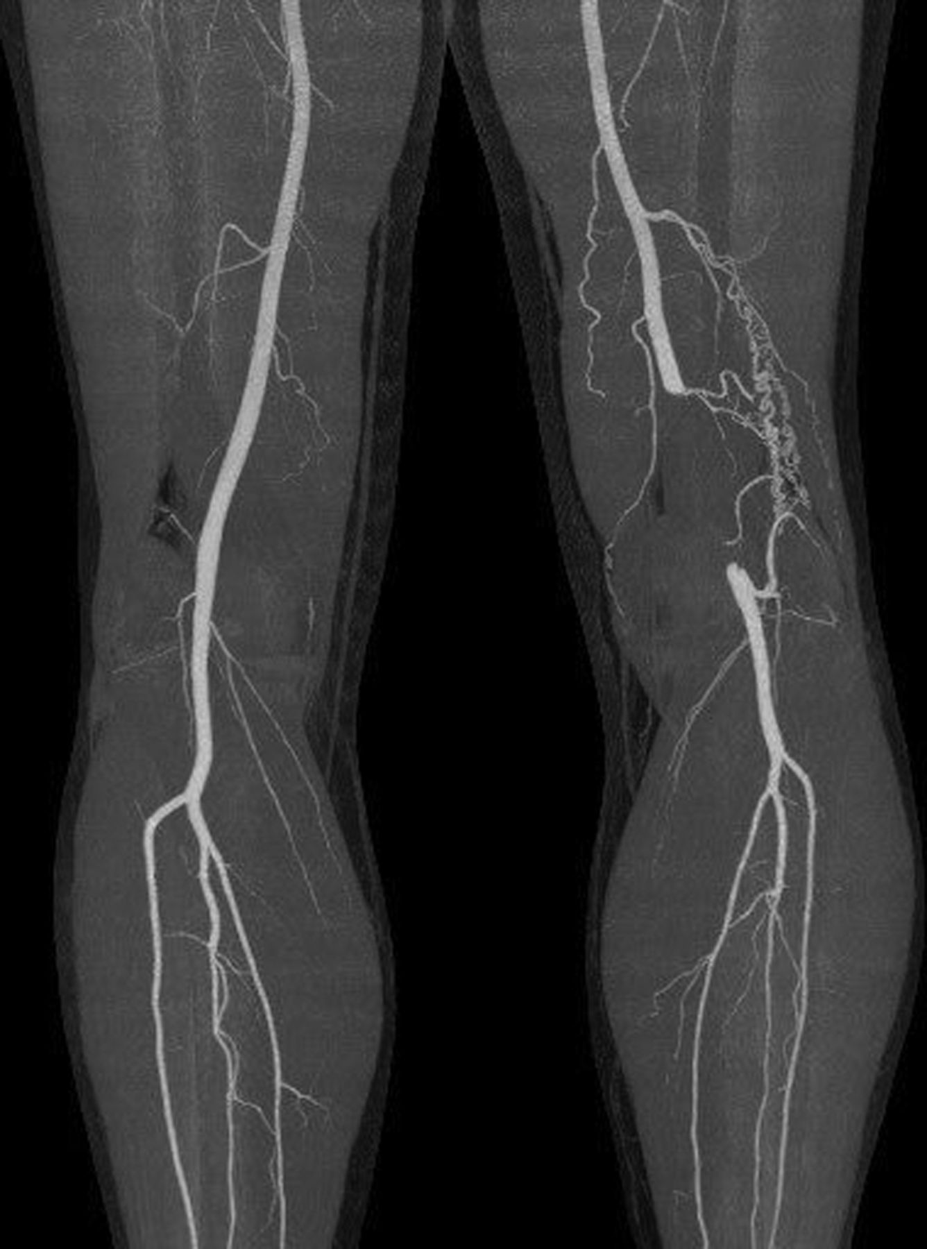
MR angiography demonstrating occlusion and significant collateralisation.
The relief was temporary, as 2 months later he presented with a recurrence of symptoms. The case was atypical as there were no significant risk factors and while the lesion was attributed to embolus, no cardiac source could be found. Therefore, the possibility of popliteal artery entrapment syndrome (PAES) was considered.
A further CT scan in the neutral and plantar flexed positions was able to demonstrate a short tight stenosis in the popliteal artery which was lying abnormally; medial rather than lateral to the medial head of gastrocnemius (figure 2). This correlated with type 2 PAES.1

CT scan demonstrating stenosis of the popliteal artery.
In theatre the patient was placed in the prone position, and an S-shaped incision was made exposing the popliteal fossa. The medial gastrocnemius ran from medial to lateral compressing the popliteal artery behind the knee in a sling fashion (figure 3). The sling was divided allowing the artery to lie free (figure 4). The SFA above the compression was hard to touch.

Popliteal artery compressed by abnormally lying medial gastrocnemius.

Popliteal artery now lying free (medial gastrocnemius released).
Three months later the patient had CT angioplasty of his SFA with immediate relief and is now symptom free.
Learning points
-
Vascular surgery atherosclerosis plays a major role in the underlying pathology in much of the field. However, anatomical abnormalities such as popliteal artery entrapment syndrome (PAES) must be considered, especially in younger patients with few or no risk factors.
-
PAES usually occurs in patients in their early 30s,1 but as demonstrated in this case it can occur in older patients.
-
CT angiography is useful in determining arterial changes and abnormalities in anatomical structures.2
-
A follow-up protocol should be maintained for PAES which may increase efficiency of treatment and subsequent investigations for further treatment if deemed necessary.
Footnotes
-
Contributors The authors of this article were directly involved in this patient care and have all contributed to the writing of the article.
-
Competing interests None.
-
Patient consent Obtained.
-
Provenance and peer review Not commissioned; externally peer reviewed.