Article Text

Statistics from Altmetric.com
Description
We describe the case of a 30-year-old woman who presented with breathing difficulty and a history of spontaneous fractures. The possibility of primary hyperparathyroidism was kept in mind and the patient was evaluated.
The initial laboratory assessment revealed the following results:
-
Parathyroid hormone (PTH) level markedly raised, PTH 2642 pmol/L (normal 1.2–5.8 pmol/L)
-
Hypercalcaemia, with corrected calcium 3.78 mmol/L (normal 2.05–2.55 mmol/L)
-
Hypophosphataemia 0.4 mg/dL (normal 0.8–1.5 mmol/L)
-
Vitamin D 62 pmol/L (normal 60–108 pmol/L)
-
Alkaline phosphatase 958 U/L (normal 30–120 U/L)
-
Haemoglobin 5.4 g/dL (normal 12–15 g/dL)
-
Total leucocyte count 8649/mL3 (normal 4500–11 000/mL3)
-
Erythrocyte sedimentation rate 14 mm/h (normal 01–22 mm/h)
-
C reactive protein 13.6 mmol/L (normal 0.76–28.5 mmol/L)
-
Random blood sugar 109 mg/dL (normal 70–120 mg/dL)
-
Blood urea 30 mg/dL (normal 20–40 mg/dL)
-
Creatinine 0.7 mg/dL (normal 0.1–1.2 mg/dL)
-
Sodium 135 mmol/L (normal 135–145 mmol/L)
-
Potassium 3.7 mmol/L (normal 3.5–5.5 mmol/L)
-
Chloride 102 mmol/L (normal 96–106 mmol/L)
-
Magnesium 2.1 mg/dL (normal 1.3–2.1 mmol/L).
-
Arterial blood gas investigations revealed partial pressure of oxygen 90 mm Hg and partial pressure of carbon dioxide 30.2 mm Hg, and pH 7.34
An ultrasound scan of the neck showed a right inferior lobe parathyroid adenoma, which was further confirmed by a Sesta MIBI scan (figures 1⇓–3).

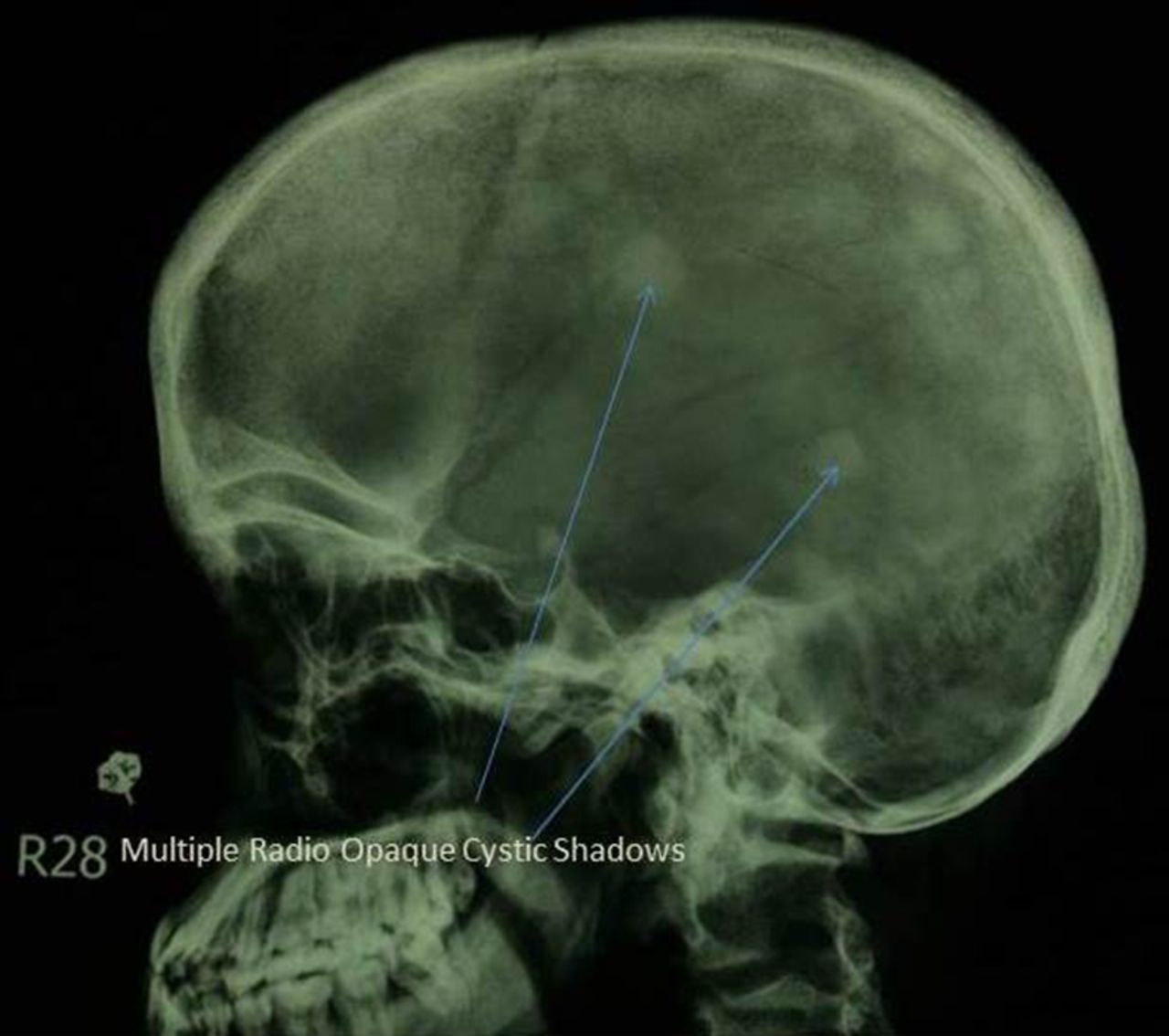
X-ray of the skull showing multiple rounded radio-opaque shadows indicating ‘moth-eaten’/cystic lesions.

X-ray of the chest showing marked thoracic cage deformity, with marked osteoporosis and pathological fractures of the upper end of each humerus. There are also multiple fractures of the ribs.

X-ray of the femur showing malunion of a mid-shaft femur fracture with callous formation and bowing.
An emergency surgery opinion was taken and the patient underwent excision of a parathyroid adenoma measuring 5.9×4.5 cm.
Following surgery, within 24 h the PTH levels dropped to a normal level (5.3 pmol/L).
Initial treatment for this patient was administration of pamidronate 60 mg as a single dose, given intravenously as a slow infusion over 12 h along with 5% dextrose normal saline (DNS) 100 mL/h infusion.
Three days after surgery, the patient developed hungry bones syndrome manifesting as symptomatic hypocalcaemia with signs and symptoms such as a positive Chvostek’s sign, carpopedal spasm and paraesthesias.1 This syndrome occurs because of rapid calcium deposition into the bone after levels of PTH have normalised, causing serum calcium levels to decrease.2 This was managed with injection of calcium gluconate 10% 10 mL every 6 h intravenously along with oral calcium carbonate and oral calcitriol. Hungry bone syndrome can be avoided by frequent monitoring of serum calcium levels and careful attention to laboratory evidence and clinical signs and symptoms of hypocalcaemia. Orthopaedic surgeons managed the femoral fractures with bilateral femoral nailing.
The patient was discharged on oral calcium carbonate 500 mg twice daily, oral calcitriol and oral alendronate.
Regular follow-ups were advised with monitoring of serum calcium and creatinine levels every 6 months along with yearly bone densitometry.3
This case highlights the importance of the early detection and management of hyperparathyroidism in order to prevent long-term orthopaedic and renal complications. The reported incidence of fractures in hyperparathyroidism is very low, about 10% in two large studies.4
The orthopaedic complications of hyperparathyroidism could have been avoided with early detection and treatment of the hyperparathyroidism.
Learning points
-
This case demonstrates that bone deformities such as moth-eaten/cystic lesions, as shown in an X-ray of the skull, and spontaneous fractures with lytic bone lesions should prompt the physician to investigate for primary hyperparathyroidism.
-
It is important to look for and recognise ‘hungry bone syndrome’ after surgical removal of a parathyroid adenoma, and to initiate prompt treatment.
-
This case also demonstrates the necessity of a team effort including input from physicians, an endocrine surgeon and an orthopaedic surgeon.
Acknowledgments
The authors would like to express their gratitude to everyone who helped in this project.
Footnotes
-
Competing interests None.
-
Patient consent Obtained.
-
Provenance and peer review Not commissioned; externally peer reviewed.