Article Text

Statistics from Altmetric.com
Description
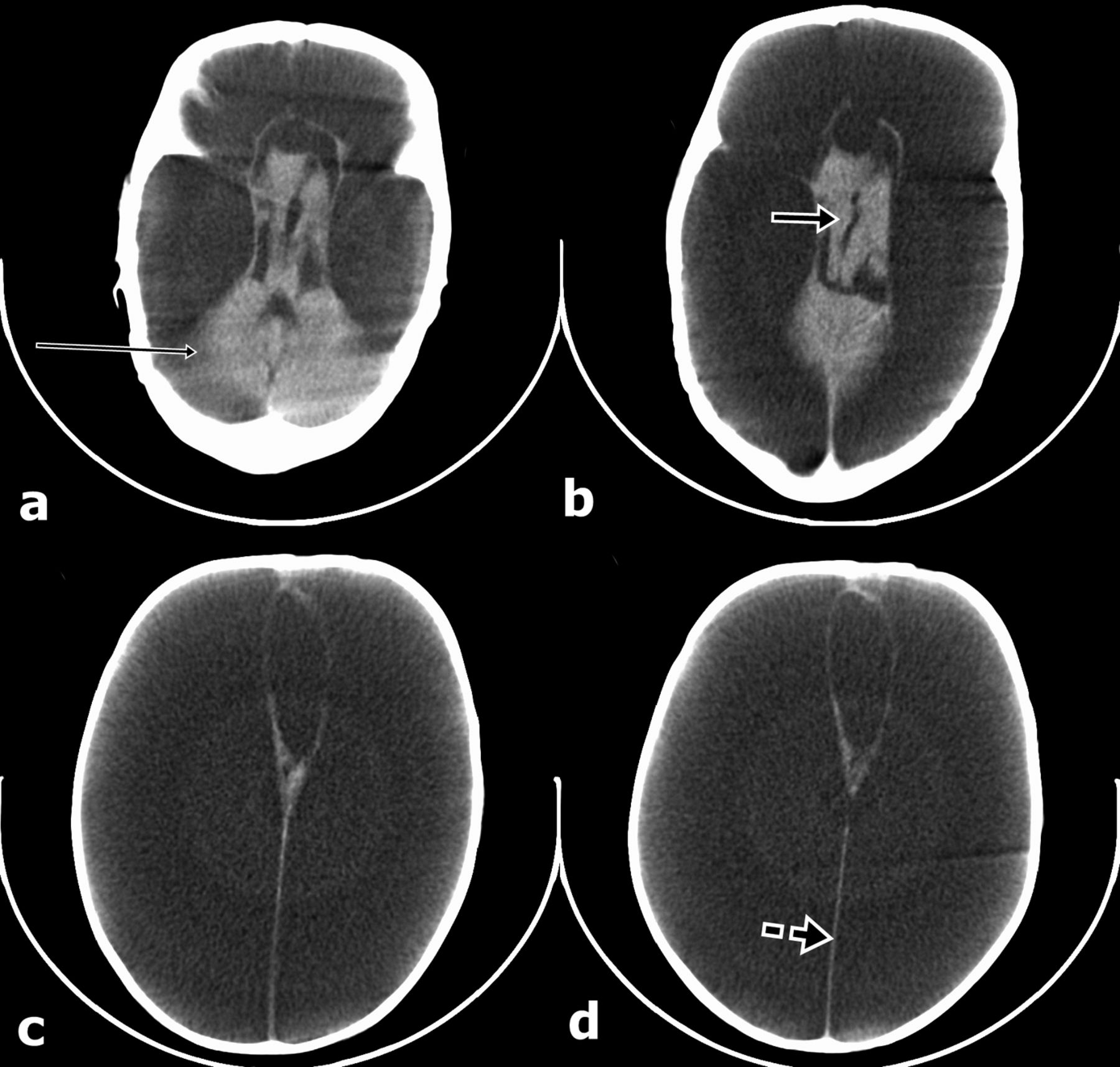
A 2-year-old female child presented with a history of delayed developmental milestones. According to her mother, the child was born out of a normal vaginal delivery and was apparently well after birth. However, by 6 months of age, the mother noticed that she was not interactive and did not recognise her. The child had failed to achieve head control by that time, but due to the ignorance and poverty of her family she did not get proper medical attention. At the time of presentation, she was still unable to recognise her mother, to speak or stand without support. She had hypertonia of all four limbs and plantar reflex was extensor bilaterally. Her fundus was normal and head appeared large with a circumference of 50 cm (>95th percentile for her age). Non-contrast CT of the child showed complete absence of bilateral cerebral hemispheres, replaced with fluid-filled spaces of cerebrospinal fluid density. Posterior fossa of the patient was normal with some soft tissue around the third ventricle representing malformed thalamic tissue. Intact falx cerebri was also seen in the interhemispheric plane, helping us to make a diagnosis of hydranencephaly (figure 1).

(A–D): Axial non-contrast CT images of 2-year-old baby taken at multiple levels showing bilateral cerebral hemispheres being replaced by the fluid containing spaces of cerobrospinal fluid density. Normal posterior fossa (thin arrow), intact falx (broken arrow) and malformed thalami (thick arrow) are seen.
Hydranencephaly is an encephaloclastic anomaly characterised by the absence and replacement of the cerebral hemispheres with cerebrospinal fluid and necrotic debris, covered by leptomeninges. Hydranencephaly is the result of destruction and resorption of preformed solid cerebral tissue commencing before birth.1 The currently accepted pathogenic mechanism is an in utero bilateral internal carotid artery obstruction with evidence that the process might begin by as early as 8–12 weeks of gestation.2 Parts of brain supplied by the posterior cerebral and vertebral arteries including the cerebellum, brain stem, thalamus and basal ganglia as well as choroids plexus are usually preserved. Hydranencephaly can be differentiated from extreme hydrocephalus by the presence of a thin rim of a cortical mantle in the latter, whereas alobar holoprosencephaly is associated with the presence of falx and frequent coexisting midline facial abnormalities.
Learning points
-
Hydranencephaly usually presents with severely delayed milesones during early childhood.
-
Head size may be enlarged despite the absence of bilateral cerebral cortex.
-
Non-contrast CT imaging is diagnostic.
Footnotes
-
Contributors AMM and AK were responsible for clinical workup and follow-up care of the patient. MA and EU were involved in diagnostic imaging. All authors contributed to the write-up and approval of the manuscript.
-
Competing interests None.
-
Patient consent Obtained.
-
Provenance and peer review Not commissioned; externally peer reviewed.