Article Text

Statistics from Altmetric.com
Description
In January 2012 a 51-year-old woman was admitted to our department for a slow increasing right hemiparesis with right brisk deep tendon reflexes. She suffered from Sjogren syndrome, fibromyalgia and hepatitis B virus (healthy carrier).
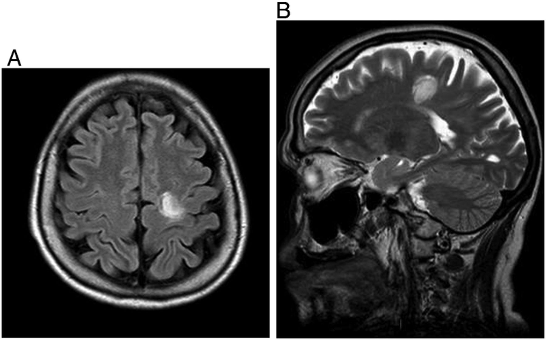
Conventional MRI scans (figure 1A,B) were typical for Balò's sclerosis.1

At diagnosis. (A) Axial fluid attenuated inversion recovery sequence and (B) sagittal T2-sequence evidence the typical concentric ring lesion in the white matter.
The patient rejected a brain biopsy. In order to better define the lesion, we therefore performed advanced radiological techniques such as diffusion, spectroscopy and perfusion.
In the diffusion sequences (figure 2A) a concentric ring of unrestricted diffusion appeared clearly distinguishable.2

At diagnosis. (A) Diffusion shows a restricted peak depicting the classical ring of demyelination and remyelination. (B) Spectroscopy shows a high ratio Cho/N-Acetylaspartate, double pick of lactate-lipids. (C) Perfusion MRI analysis, based on four regions of interest depicted in D (A, pink, B, white, C, blue, D, red) suggests that the central layers of the ring are more perfused compared to the peripheral ones.
The spectroscopic image (figure 2B) of the lesion documented increased choline peak and decreased N-acetyl aspartate peak, but normal peaks in the normal appearing white matter near the lesion.1
The perfusion analysis (figure 2C), based on four different regions of interest (figure 2D), suggested a decreasing gradient of perfusion from the centre to the periphery of the lesion, supporting the hypothesis that the centre of the ring corresponds anatomically to a deep venous vessel.3 Authors are not aware of previous report of perfusion studies in Balò's sclerosis.
Cerebrospinal fluid analysis detected oligoclonal bands, without anti-aquaporin-4 antibodies.
High-dose intravenous methylprednisolone was started (1 g/day for 10 days) and followed by oral prednisone (1 mg/kg/day for 2 months, then slowly tapered in 2 months) with concomitant lamivudine as antiviral prophylaxis. The right paresis fully recovered after 2 weeks and has not relapsed after 11 months.
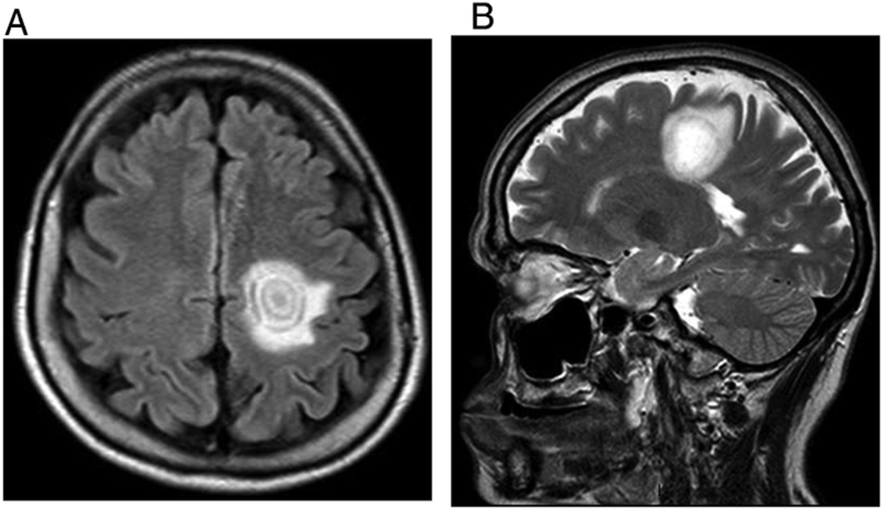
The 8 months follow-up MRI showed a reduction in the lesion volume and absence of new demyelinating lesions (figure 3A,B).
At 8 months follow-up (after treatment). (A) Axial fluid attenuated inversion recovery sequence and (B) sagittal T2-sequence evidence the reduction in the lesion volume compared to figure 1.
Learning points
-
Balò's concentric sclerosis is a rare demyelinating disease presenting with a concentric ring in the white matter. The clinical and radiological features can mimic other diseases such as primary central nervous system lymphoma, low-grade glioma or stroke.
-
A brain biopsy should be obtained whenever possible. However, nowadays advanced neuroimaging studies (spectroscopy, MRI diffusion and perfusion) seem to be a reliable tool for the diagnosis.
-
Balò's sclerosis could respond very well to high-dose steroids alone or in combination with other immunosuppressive treatments (eg, plasma exchange).
Footnotes
-
Competing interests None.
-
Patient consent Obtained.
-
Provenance and peer review Not commissioned; externally peer reviewed.