Article Text

Statistics from Altmetric.com
Description
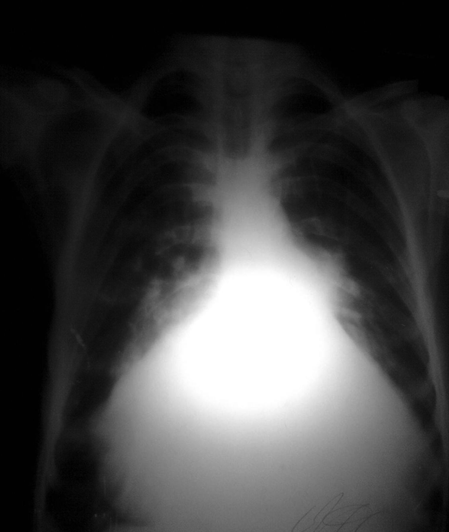
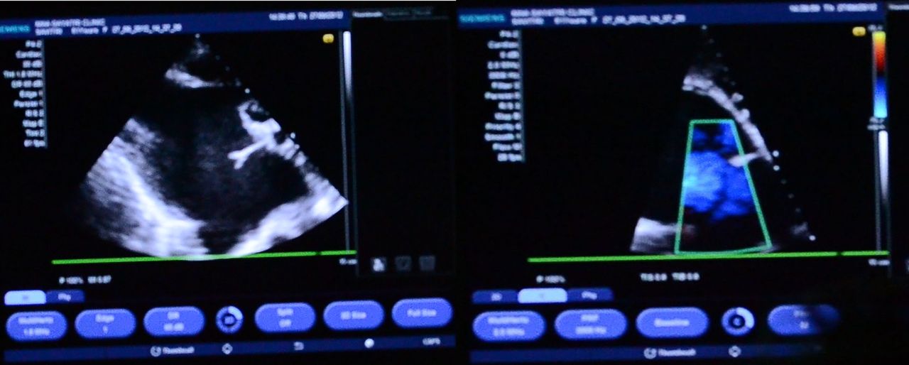
A case of prominent jugular venous pulse (JVP) with a clear wave pattern and the echocardiography of the condition responsible is discussed. A 63-year-old woman living in a hilly region presented to us with a 20-day history of orthopnoea and swellings in her legs and a 1-year history of exertional dyspnoea and palpitations. She had a 1-year history of New York Heart Association (NYHA) class III cardiac failure and was being treated at a local hospital with diuretics for dilated cardiomyopathy. For the last 15 days she was finding it difficult to walk up hills. On physical examination, she had an elevated JVP with visible large ‘a’ and ‘v’ waves with fusion, which was prominent even in the sitting position (video 1), and a displaced apex beat in the sixth intercostal space. She had a left parasternal heave and a murmur caused by tricuspid regurgitation. She had a wide and fixed S2. Her chest x-ray revealed cardiomegaly (figure 1) and 2D echocardiography showed a large atrial septal defect with significant left to right shunt and pulmonary hypertension with the ‘matchstick’ sign (figure 2).

Chest x-ray showing cardiomegaly.

(A,B) Echocardiography showing the ‘matchstick’ sign with blood gushing from the left to right atrium.
Video demonstration of a prominent jugular venous pulse
The ‘v’ wave reflects the passive increase in pressure and volume of the right atrium as it fills in late systole and early diastole. Normally the crests of the ‘a’ and ‘v’ waves are approximately equal in amplitude.1 In patients with an atrial septal defect, the higher left atrial pressure is transmitted to the right atrium and ‘a’ and ‘v’ waves are equal or the ‘v’ wave is larger than the ‘a’ wave.2 Other conditions producing a prominent JVP are tricuspid regurgitation and constrictive pericarditis.3
Learning points
-
A raised jugular venous pulse with ‘v’ waves of higher amplitude than the ‘a’ waves can be seen in conditions such as atrial septal defect, tricuspid regurgitation and constrictive pericarditis.
-
The collapse in neck veins is the most dramatic movement and should be timed when determining the type of wave.
-
Prominent jugular venous pulse in an adult with symptomatic ASD denotes severe pulmonary hypertension.
References
Footnotes
-
Competing interests None.
-
Patient consent Obtained.
-
Provenance and peer review Not commissioned; externally peer reviewed.