Article Text

Statistics from Altmetric.com
Description
A 48- year-old man presented with predominant nocturnal cough with minimal sputum, since 2 years. He had frequent retrosternal pain with occasional lower dysphagia since 6 months. He had no weight loss, fever or wheeze. He was a hypertensive for 9 years. He had left leg fracture at the age of 11 and developed permanent limb shortening in spite of multiple surgeries. He had mild cognitive defect since 1 year.
His chest x-ray (figure 1) showed a large retro-cardiac globular shadow without a fluid level, unclear borders of left hemi diaphragm and multiple pulmonary nodules. The radiological diagnosis given by the radiologist was a posterior mediastinal mass with multiple lung metastases, most likely a lower oesophageal malignancy.
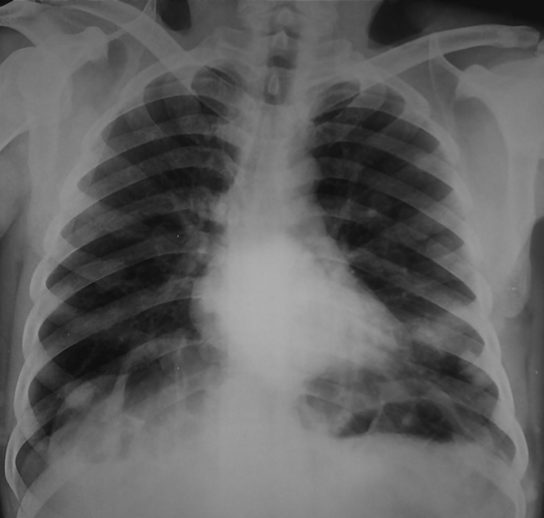
Chest x-ray posteroanterior view.
The reported large mediastinal mass and metastases were actually the tumours on his chest wall (figures 2 and 3). The patient is suffering from Neurofibromatosis type I (NF1), also known as Von Recklinghausen disease.1 The chest radiogram was reported by a radiologist, who neither saw the patient personally nor enquired into the patient's history. Note the multiple café au lait spots on the patient's skin, another characteristic of this disease.2 Cognitive defects, peudoarthrosis of tibia and long bone dyplasia are also known to occur in NF1.3 Pheochromocytoma was not present in him but is known in NF1.
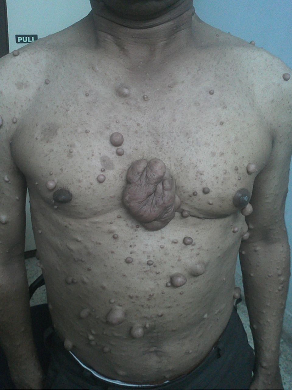
Large chest wall tumour on the lower end of sternum.

Posterior chest wall tumours.
The patient underwent oesophago-gastroscopy which confirmed the presence of GERD (Gastro-oesophageal reflux disease). Oesophageal malignancy or hiatus hernia was ruled out. The cough responded to antireflux treatment within 1 month.
Learning points
-
Soft tissue lesions on the anterior or posterior chest wall can be easily mistaken as intrapulmonary lesions on a chest radiogram posteroanterior (PA) view.
-
A standard chest radiogram PA view should always cover the lateral chest wall. Had this been done, the skin nodules would have been easily picked up.
-
A clinician should always clinically correlate an x-ray and its report, before making a diagnosis.
Footnotes
-
Competing interests None.
-
Patient consent Obtained.
-
Provenance and peer review Not commissioned; externally peer reviewed.