Article Text

Statistics from Altmetric.com
Description
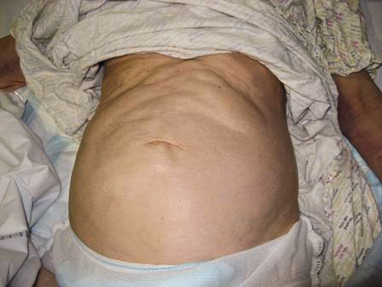
A 62-year-old female presented with a 3-week history of non-specific abdominal pain. She had a history of rectal prolapse and was awaiting rectopexy. Clinical examination revealed gross anterior abdominal wall surgical emphysema (figures 1 and 2) as well as surgical emphysema of the pararectal region on digital rectal examination. There were no signs of peritonitis. A rigid sigmoidoscopy on admission revealed no obvious mucosal abnormality. There was no history of any rectal trauma. An erect chest radiograph confirmed a pneumoperitoneum. A CT scan, performed to identify the site of perforation, revealed a thickened rectosigmoid surrounded by large amounts of free air (figure 3) with extensive surgical emphysema extending to the chest wall and groin. There were large amounts of free air within the retroperitoneal space and around the aorta (figure 4). A diverting sigmoid loop-colostomy was fashioned to allow healing of the rectosigmoid perforation. The patient’s postoperative period was unremarkable and she was discharged 1 week later. Spontaneous rectal perforation is an extremely rare condition with only 65 reported cases.1 2 The majority of reported cases are either iatrogenic or associated with rectal prolapse.3 4 Other causes of rectal perforation include malignancy, diverticular disease, stercoral ulceration, trauma and ulcerative colitis. Constipation and rectal prolapse may lead to stercoral ulceration due to pressure necrosis from fecolomas causing perforation.5 A retroperitoneal perforation can make diagnosis difficult due to the lack of symptoms.6 Clinical examination and imaging with CT can help with diagnosis and subsequent management.

Anterior wall surgical emphysema.
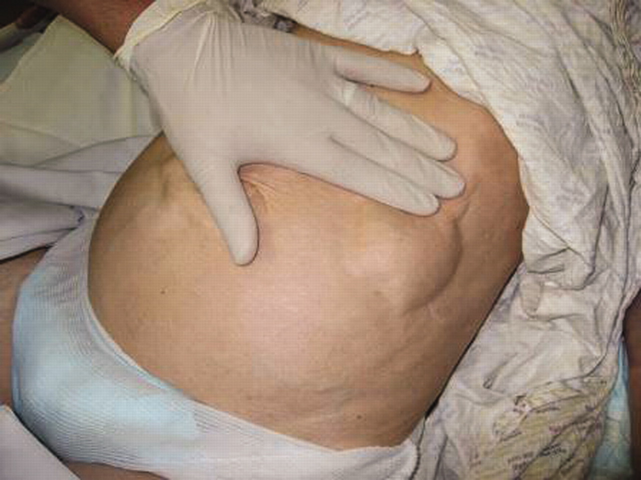
Surgical emphysema prominent on palpation.

(a) Extensive surgical emphysema in abdominal wall. (b) Thicken rectosigmoid with surrounding air in the pararectal region.

(c) Retroperitoneal free air extending into thorax and around the aorta. (d) Free intra-abdominal fluid.
Footnotes
-
Competing interests None.
-
Patient consent Obtained.