Article Text

Statistics from Altmetric.com
Description
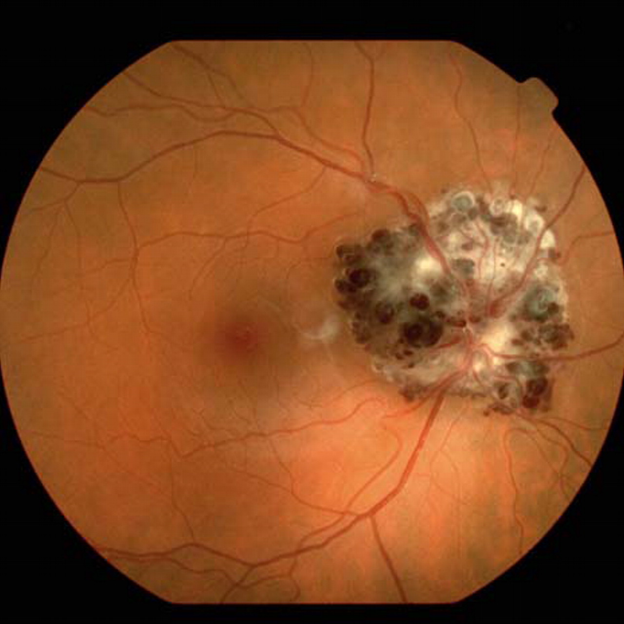
A healthy 29-year-old woman presented at 20 weeks’ gestation with a 3-week history of a paracentral scotoma in her right eye. Visual acuity was 20/20 in both eyes. Fundus examination of the right eye showed a mass of numerous, grape-like, aneurysms filled with dark blood along the optic disc. White fibroglial tissue covered the surface. A triangular haemorrhage was observed in the macular area, under the internal limiting membrane, and stopping just at the border of the fovea (figure 1). Humphrey visual field testing showed an enlarged blind spot (figure 2). MRI revealed no additional abnormalities. The haemorrhage resolved favourably in 1 month (figures 3 and 4).

Fundus photograph of the right eye showing a mass of dark saccular aneurysms with overlying white fibroglial tissue. Note the triangular subinternal limiting membrane along the temporal margin of the lesion that stops near the fovea. Another subretinal haemorrhage can be seen in the upper margin.

Humphrey visual field testing showing an enlarged blind spot.
Clinical appearance showed resolution of the haemorrhage at 1 month.

Late-frame of fluorescein angiogram demonstrated incomplete perfusion of the lesion and typical plasma-erythrocyte sedimentation.
Cavernous haemangioma of the optic disc is an infrequent vascular haemartoma and it is almost always asymptomatic.1 2 Growth is rare although enlargement of the fibroglial component and fluctuations in the size of the lesion have been described.1 Bleeding is infrequent. Most haemorrhages are minimal and not associated with long-term loss of visual acuity.1
We present a case of an extremely large cavernous haemangioma of the optic disc diagnosed during pregnancy following a subinternal limiting membrane haemorrhage in the macular region. Recently, Patikulsila et al reported a similar case in a 15-year-old boy in whom the lesion remained stable without haemorrhages.2 Smith et al described a case of a retinal cavernous haemangioma that haemorrhaged during labour. They presumed that venous pressure elevation during Valsalva manoeuvre broke the wall of the aneurysms.3
It is unclear whether the haemorrhage was related to the pregnancy in our patient as no Valsalva stress was identified. Location of the haemorrhage was critical to diagnosing the tumour. The bleeding caused visual disturbance but loss of vision was not significant.
Footnotes
-
Competing interests None.
-
Patient consent Obtained.