Article Text

Summary
The authors report a case of a 41-year-old woman who was admitted to the emergency department of our hospital because of acute right flank pain. Laboratory investigations and cultures were negative. A transabdominal ultrasonography revealed a large mass of the upper pole of the right kidney as an incidental finding.
Statistics from Altmetric.com
Case presentation
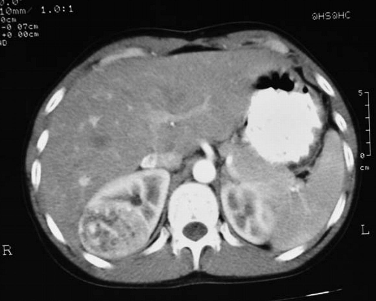
A 41-year-old woman was admitted to the emergency department of our hospital because of acute right flank pain. Laboratory investigations and cultures were negative. A transabdominal colour doppler ultrasonography scan using head transducer 1.75–4 MHz (Siemens/Acuson; Aspen, Mountain view, California, USA) revealed a large (5×4 cm), highly hyperechoic well-demarcated exophytic mass with shadowing, of the upper pole of the right kidney as an incidental finding (figure 1). Subsequently abdominal CT spiral scan (10/10 mm sections GE SX Prospeed before and after contrast injection) showed a mass with mixed hyperdense and fatty tissue elements (figures 2, 3). Renal excretion and filling of the urinary bladder were both satisfactory. There was no evidence of lymph node enlargement or other space occupying lesion in the abdomen. All the diagnostic imaging findings were consistent with the diagnosis of angiomyolipoma. A follow-up was recommended.

Large uniformly hyperechoic mass of the upper pole of the right kidney with sharp borders where echogenicity of the mass is at least equal to that of renal sinus fat. A picture of the classic ultrasonography appearance of acute myeloid leukaemia.

CT shows well-demarcated mass of the upper pole of the right kidney with mixed soft tissue and fatty tissue elements.
CT picture of angiomyolipoma. Enhancement of smooth muscle and vascular portions of the tumour after administration of contrast.
Discussion
Angiomyolipoma is a benign renal neoplasm composed of fat, vascular and smooth muscle elements. It has a prevalence of about 0.3–3%. Two types are described: isolated angiomyolipoma and angiomyolipoma associated with tuberous sclerosis. Isolated angiomyolipoma occurs sporadically, is often solitary and accounts for 80% of all angiomyolipomas.1 Shadowing on sonography is seen in 33% of acute myeloid leukaemias.2 3 In the general population in both sexes angiomyolipoma are most common in the age group 40–45 years. The mean age at presentation in patients with isolated angiomyolipoma is 43 years. This neoplasm is about four times more common in women than in men and interestingly 80% of cases involve the right kidney. Angiomyolipomas have thicker arteries than normal but abnormally weak vessel walls which predispose to aneurysm formation.4 Angiomyolipoma that is associated with tuberous sclerosis accounts for 20% of angiomyolipomas. The lesions are typically larger than isolated angiomyolipomas, and they are often bilateral and multiple. Angiomyolipomas occur in 80% of patients with tuberous sclerosis.1 The male-to-female distribution of angiomyolipoma in patients with tuberous sclerosis is almost equal. Angiomyolipomas occur in young women with lymphangiomyomatosis without other stigmata of tuberous sclerosis. Although angiomyolipomas are considered benign, rare cases that are possibly related to multicentric disease have been reported regarding extension into the renal vein, the inferior vena cava, or both; deposits in the regional lymph nodes have also been reported. The risk is probably very low in tumours that are <3 cm in diameter. Apart from this group with tuberous sclerosis, follow-up may be reasonably restricted to patients with sporadic tumours >4 cm in diameter, in whom the prevalence rate of haemorrhagic complications is higher. Thin section multidetector CT is the preferred method to demonstrate fat in problem cases (table 1).5
Examination methods of angiomyolipoma
Footnotes
-
Competing interests None.
-
Patient consent Obtained.