Article Text

Statistics from Altmetric.com
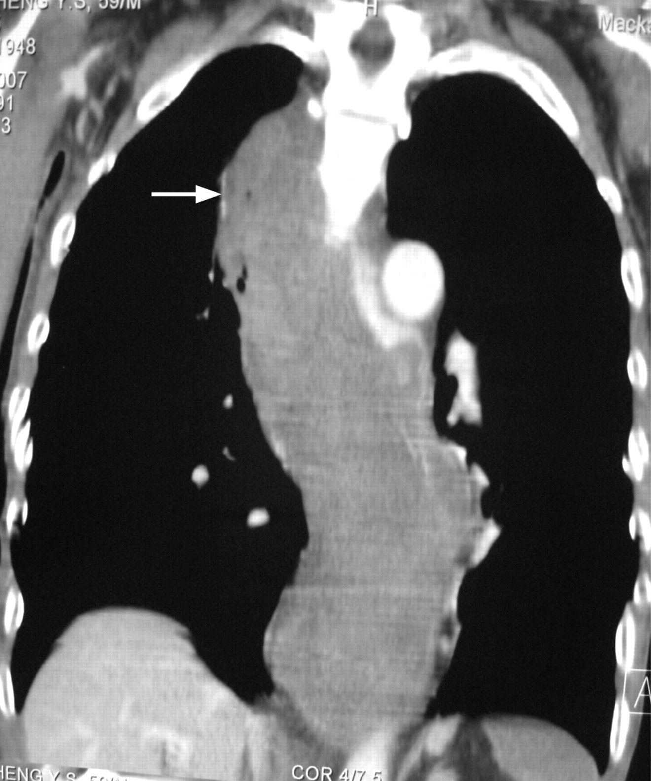
A 59-year-old man vomited coffee ground material for 2 days. He had had oesophageal cancer surgically removed 7 months previously followed by gastric tube reconstruction. An initial chest radiograph showed no significant mediastinal lesion (fig 1). However, the patient suddenly began to vomit fresh blood and became hypotensive. A repeat chest radiograph revealed acute widening of the mediastinum (fig 2). Aortic dissection or osophageal perforation (reconstructive gastric tube) were suspected. However, a chest CT scan showed that the reconstructed gastric tube was markedly distended by blood (fig 3). There was no evidence of aortic dissection or perforation.


Acute mediastinal widening on chest radiography may be found in acute aortic dissection or oesophageal perforation with mediastinitis.1 In our patient the normal anatomy had been disrupted by the surgery, allowing the reconstructed tube to expand in a way the normal oesophagus would not have. Interestingly, when the tube was empty the mediastinum appeared normal. It was only acute distension with retained blood that caused the alarming radiographic appearance.
Acknowledgments
This article has been adapted from Chang C-W, Chang W-H, Shih S-C, Tsai S-J. Acute widening of mediastinum in a patient with oesophageal cancer after surgery Emergency Medicine Journal 2008;25:382
REFERENCE
Footnotes
Competing interests: None.
Patient consent: Patient consent has been received for publication of the details of this case.