Article Text

Statistics from Altmetric.com
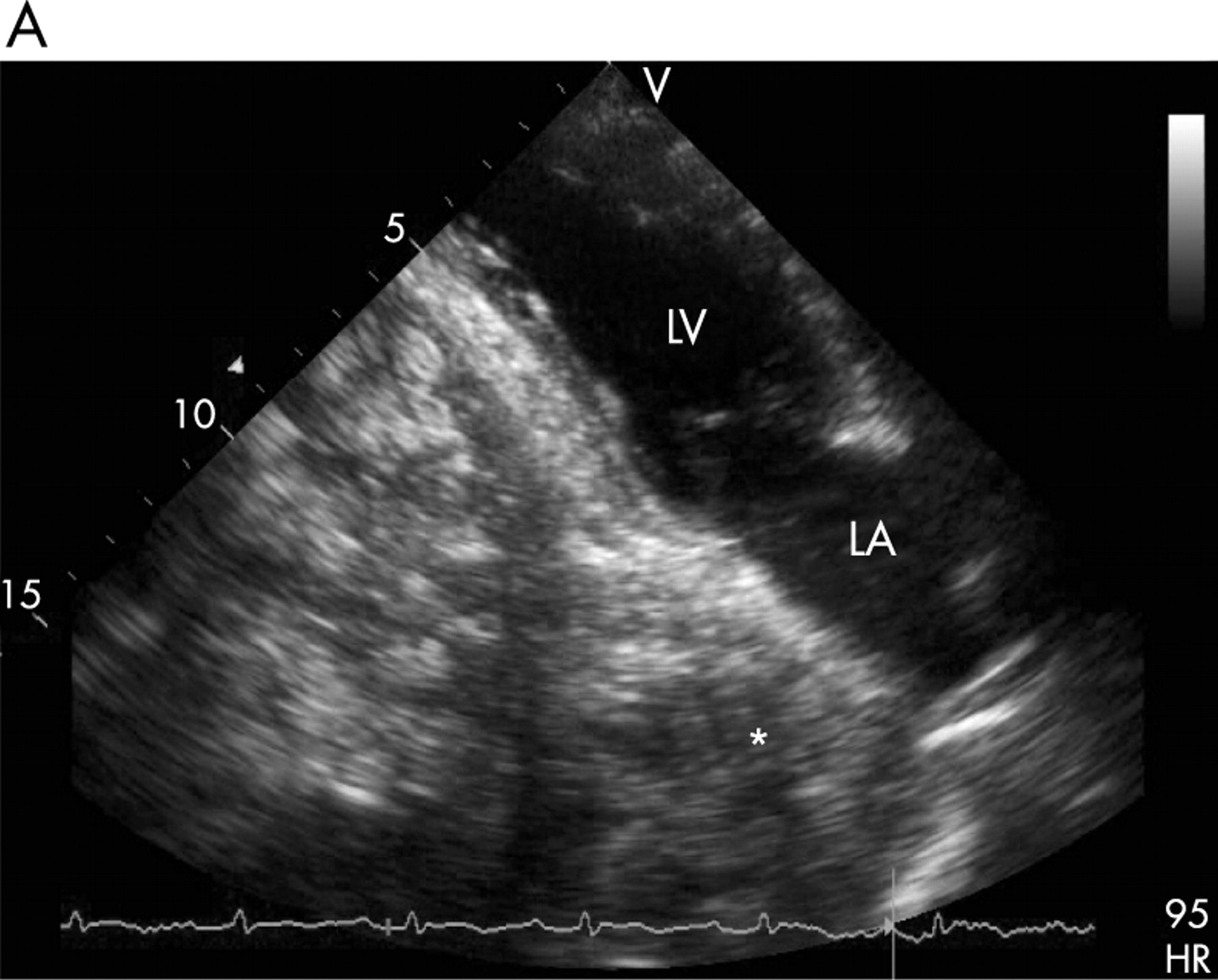
An 85-year-old woman was examined at the echocardiography laboratory during a predischarge examination after an uncomplicated, circumscribed acute myocardial infarction.
An undefined liquid-containing mass (*) was observed behind the left atrium (LA), with no evidence of compression (panel A). This mass could be clearly demonstrated (arrow) after ingestion of 300 ml sparkling water mixed with an ampoule of the echo contrast medium Echovist 300 (d-galactose suspension; Schering AG, Berlin, Germany), and the performance of a Valsalva manoeuvre (panel B). The video recording of the examination shows a dynamic filling of the mass behind the LA during the abdominal compression manoeuvre, and its emptying towards the stomach after relaxation. The patient showed no symptoms at all during the whole examination.
A hiatal hernia masked as an LA mass can be diagnosed accidentally during echocardiographic examination. Most patients are asymptomatic, whereas a few patients present symptoms of gastro-oesophageal reflux. Arrhythmias and heart failure have been reported. This patient presented no history of recurrent gastrointestinal symptoms or postprandial syncope, therefore, conservative treatment was recommended.
To view video footage visit the Heart website—http://heart.bmj.com/supplemental


Acknowledgments
This article has been adapted from Buss S, Katus H A, Mereles D. Dynamic changing mass behind the left atrium Heart 2007;93:1583