Article Text

Statistics from Altmetric.com
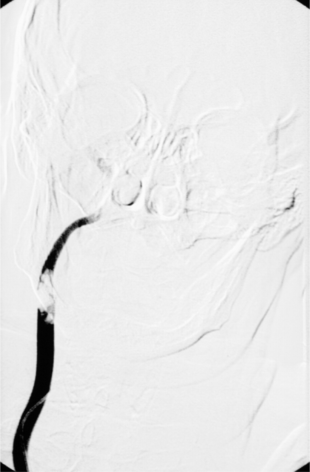
A 56-year-old woman presented with fluctuating left-sided hemiparesis. The initial computer tomography scan showed signs of a frontal cortical ischaemia within the territory of the prerolandic artery. Duplex sonography showed a pulsating structure within the right internal carotid artery. An angiographic investigation showed a large polymorphic thrombus within the internal carotid artery (fig 1). Atrial fibrillation was detected in the electrocardiogram. The echocardiogram showed dilation of the left atrium. The heart valves were intact and no cardiac thrombi could be found.

Before a thrombectomy could be performed, the carotid artery became desobliterated spontaneously and further ischaemic lesions developed in the territories of the right arteria cerebri media and arteria cerebri anterior.
Absolute arrhythmia is the most common cause of cerebral thromboembolism.1 The intracerebral arteries are usually affected.2 In contrast, the finding of a large thrombus in the carotid artery as in the present case is very rare.3
Acknowledgments
This article has been adapted from Surov Alexey, Behrmann Curd, Kornhuber Malte. Carotid embolism Journal of Neurology, Neurosurgery and Psychiatry 2007;78:564
Footnotes
Competing interests: None declared.