Article Text

Statistics from Altmetric.com
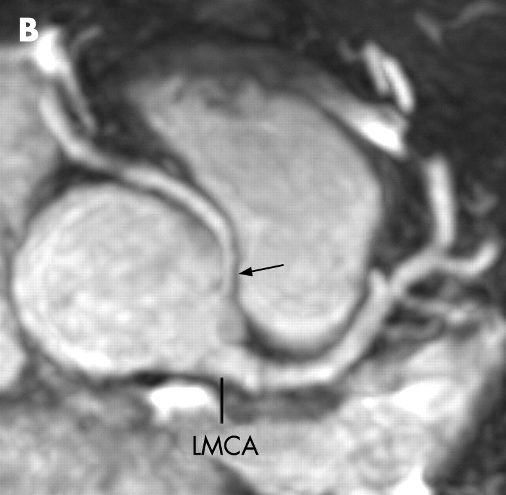
A 46-year-old man without a history of cardiac disease presented with atypical chest pain in the early morning. A 12-lead, 24-hour ambulatory electrocardiogram disclosed an ST-segment elevation in leads II, III and aVF, suggestive of vasospasm of the right coronary artery. Myocardial perfusion single-photon emission computed tomography was normal. Navigator-guided, free-breathing whole heart MRI demonstrated the right coronary artery arising from the left sinus of Valsalva separately from the left main coronary artery and coursing between the ascending aorta and the pulmonary artery (panel A). On an axial image, there was an acute angle take-off of the right coronary artery from the aorta (panel B). A curved maximum intensity projection image showed no significant stenosis in the right coronary artery (panel C). The patient was diagnosed as having vasospastic angina in the anomalous right coronary artery and treatment with calcium channel blocker and isosorbide dinitrate was started. He has had no pain for the 2-month follow-up period. To the best of our knowledge, this is the first report demonstrating three-dimensional whole heart MRI of the anomalous origin of the right coronary artery.


Acknowledgments
This article has been adapted from Sato Y, Matsumoto N, Saito S. Three-dimensional whole heart magnetic resonance imaging of anomalous origin of the right coronary artery from the left sinus of Valsalva Heart 2007;93:907