Article Text

Statistics from Altmetric.com
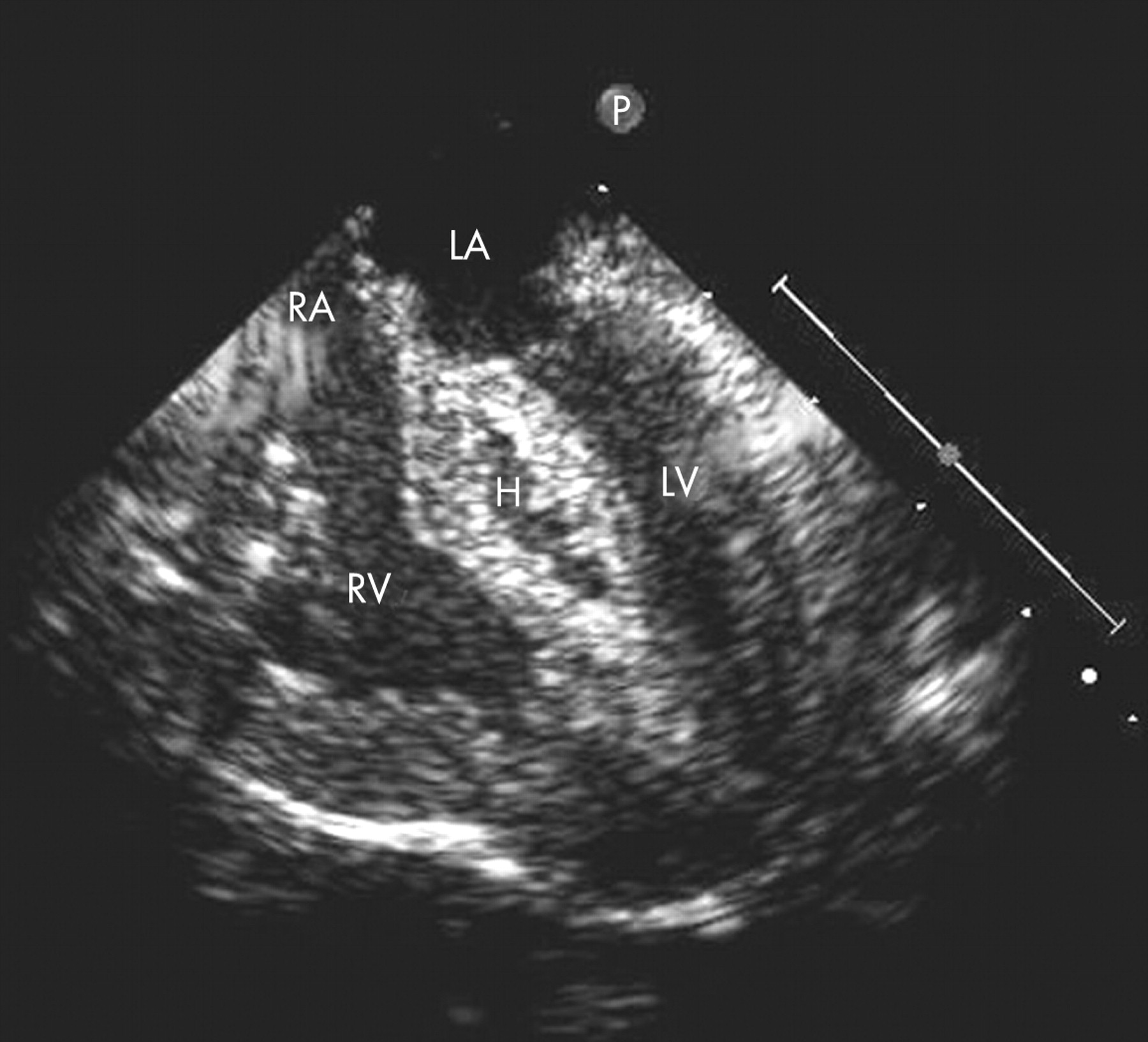
A 1-day-old boy was referred to our institution with cyanosis and respiratory distress. The chest radiograph demonstrated cardiomegaly and increased pulmonary vascularity. A cross-sectional echocardiogram disclosed obstructed mixed total anomalous pulmonary venous connections with a large secundum atrial septal defect and enlarged right cardiac chambers. The patient underwent surgical correction at 2 weeks of age. The intraoperative transoesophageal echocardiogram showed poor biventricular function along with a large hypoechogenic mass in the basal inferior septum (panel). A diagnosis of spontaneous intraventricular haematoma was made. The child left the operating room on extracorporeal membrane oxygenation (ECMO) support. Serial transthoracic echocardiograms showed progressive improvement of the ventricular function, and complete spontaneous resolution of the haematoma was noted 11 days after surgery. ECMO was discontinued on postoperative day 4 and the child was discharged home 1 month after surgery. Intraoperative spontaneous haematoma is an uncommon finding. In our experience, expectant management is warranted.
Acknowledgments
This article has been adapted from Bernasconi A, Cavalle-Garrido T, Redington A. Spontaneous intraoperative ventricular haematoma in a neonate Heart 2007;93:898