Article Text

Statistics from Altmetric.com
A 34-year-old man was admitted to our hospital (Niigata University Medical and Dental Hospital, Niigata, Japan) with dyspnoea on exertion and pretibial oedema. The chest x ray showed cardiomegaly, pulmonary congestion and right pleural effusion. The echocardiogram showed enlargement and diffuse hypokinesis of the left ventricle. Cardiac enzyme levels were not raised on laboratory examination. Symptoms were alleviated with intravenous diuretics and catecholamines, then stabilised with oral diuretics, digoxin and enalapril.
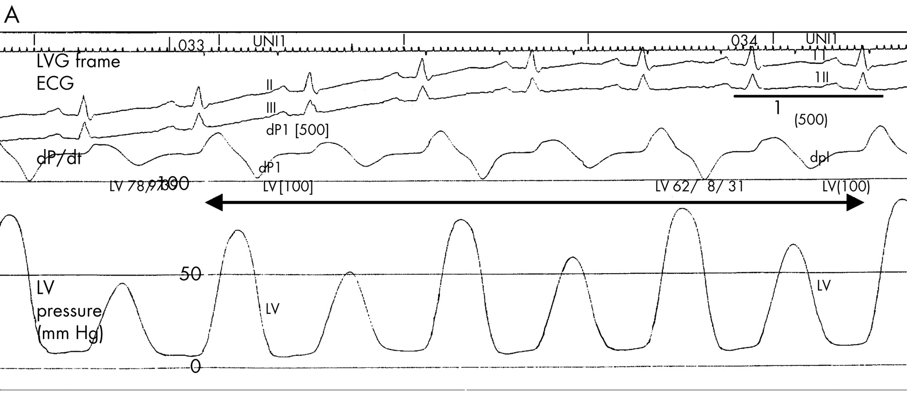
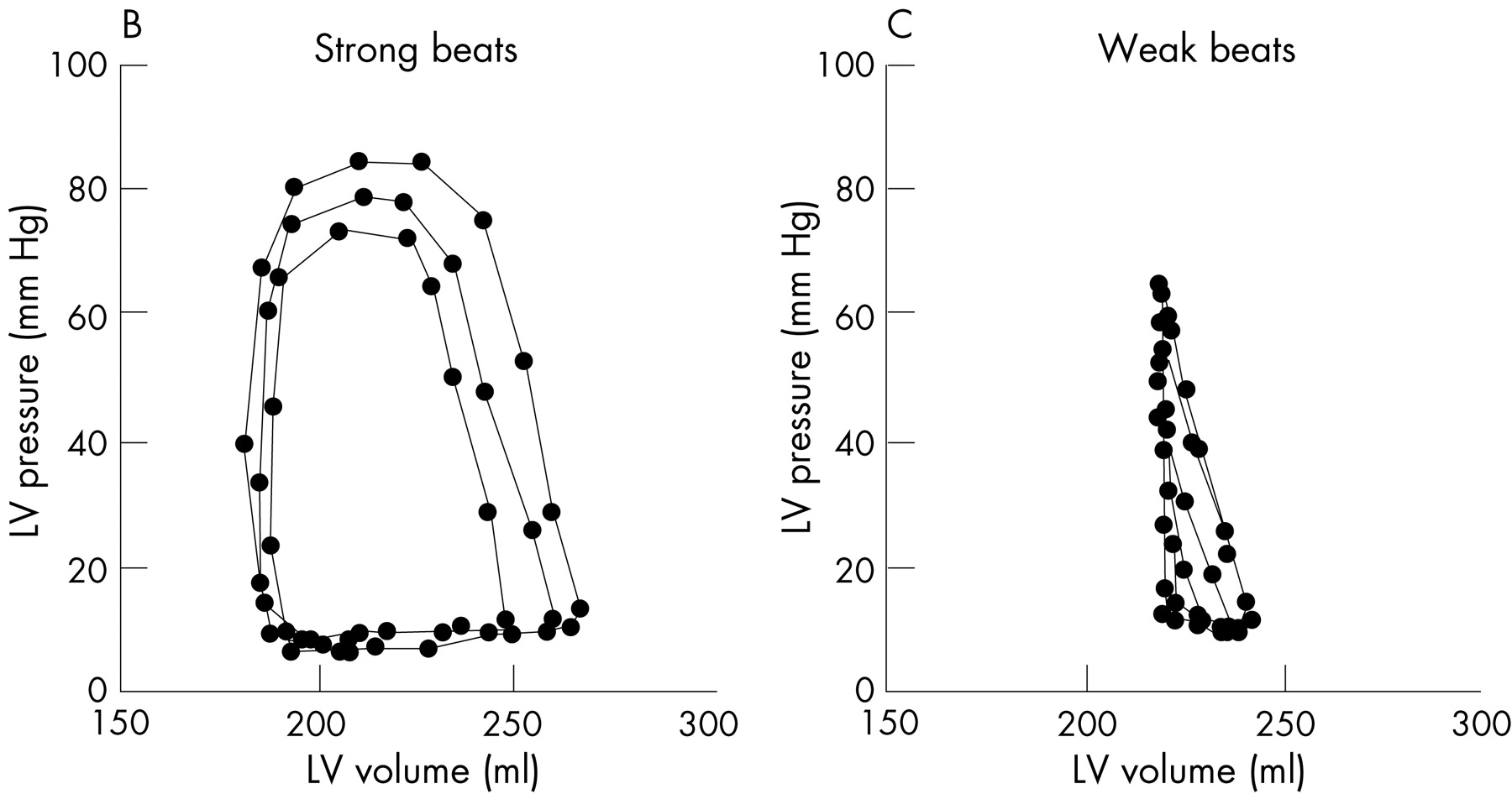
Cardiac catheterisation was performed about 1 month after admission. Coronary arteries were normal, and an endomyocardial biopsy specimen did not show inflammation or any specific findings. The patient was diagnosed with idiopathic dilated cardiomyopathy. Left ventriculography was performed and the left ventricular pressure was simultaneously recorded using a micromanometer-tipped pigtail catheter. Apparent mechanical alternans was recorded in the pressure wave form (panel A) and visualised on left ventriculography. Left ventricular volume was calculated by the Kennedy’s regression equation, and the pressure–volume loops of six consecutive beats (panel A, bidirectional arrow) were drawn. Panels B and C show the loops of three strong and three weak beats separately. The end-systolic pressure–volume points of strong beats were higher in pressure and smaller in volume than those of weak beats, suggesting that the strong and weak beats were associated with differences in contractility.
The alternating contractility during mechanical alternans was shown by drawing the left ventricular pressure–volume loops of the isolated canine heart. This is the first report of human left ventricular pressure–volume loops during mechanical alternans.


Acknowledgments
This article has been adapted from Kashimura T, Kodama M, Aizawa Y. Left ventricular pressure–volume loops during mechanical alternans in a patient with dilated cardiomyopathy Heart 2007;93:151