Article Text

Statistics from Altmetric.com
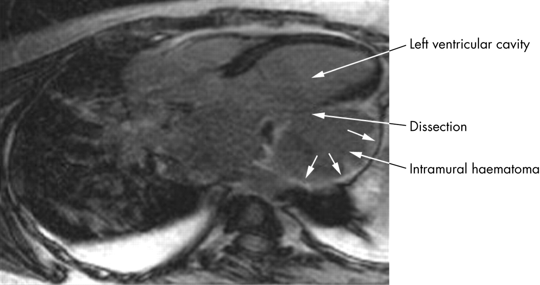
An “aneurysm” of the left ventricular (LV) free wall was found incidentally on the chest computed tomographic (CT) scan of a 39-year-old female patient. The CT scan was ordered to evaluate pneumonia refractory to antibiotics. The patient was a smoker and reported an episode of back pain a few months previously. Cardiac magnetic resonance (CMR) imaging was performed to further characterise the “aneurysm”. The three-chamber view cine (to view video footage visit the Heart website—http://www.heartjnl.com/supplemental) shows an aneurysmal formation with turbulent pulsatile blood flow from the LV cavity. The CMR delayed contrast enhanced image (see panel) shows contrast enhancement in the lateral wall of the “aneurysm” indicating infarcted myocardium. No pericardial effusion was seen. The findings were suggestive of a dissecting haematoma of the LV free wall. The coronary angiogram showed an occluded left circumflex artery as possible underlying cause for the dissection. During surgery the diagnosis of a dissecting haematoma was confirmed. Ultimately, the patient recovered and was back at work six months after the surgery.
Rupture of the LV free wall is associated with a high mortality. Subacute ruptures are less severe and the incidence of survival is greater. Intramyocardial haematoma is a type IV rupture most commonly occurring in the anterior and lateral wall of the LV. Typically the diagnosis of an intramyocardial haematoma requires surgery or is determined at autopsy. In our patient CMR delayed contrast enhanced imaging of the heart demonstrated that the “aneurysmal formation” was lined by infarcted myocardium. In concert with the lack of pericardial effusion, this suggests an intramyocardial haematoma.
To view video footage visit the Heart website—http://www.heartjnl.com/supplemental
Acknowledgments
This article has been adapted from Muehling O M, Huber A, Schmoeckel M, Behr J. Non-invasive diagnosis of an intramyocardial dissecting haematoma of the left ventricular free wall by cardiac magnetic resonance Heart 2007;93:71
Footnotes
All authors declare that there are no relevant competing interests