Article Text

Statistics from Altmetric.com
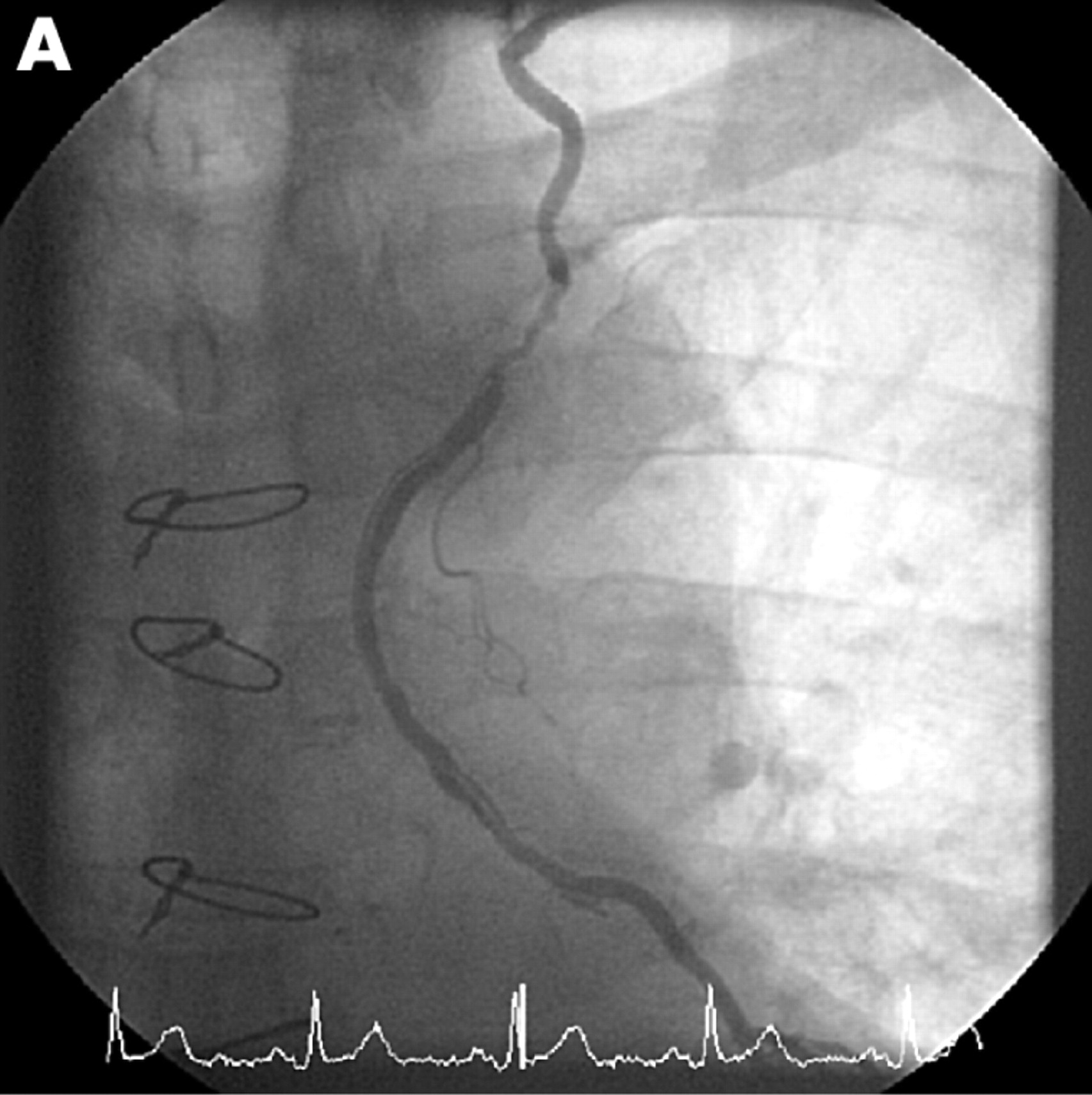
A 75-year-old man was admitted with sudden onset central chest pain, which was provoked by lifting a heavy bookcase. He had been asymptomatic since coronary artery bypass grafting (CABG) eight years previously. This included left internal mammary artery (LIMA) graft to his left anterior descending artery (LAD) and saphenous vain grafts to the obtuse marginal artery (OM) and right coronary artery (RCA). His cardiac risk factors were type 2 diabetes mellitus and a family history of premature coronary disease. Clinical examination was unremarkable and the ECG showed T wave inversion in V1–V3. Troponin T was elevated at 2.8 u/l (normal < 0.01 u/l). Coronary angiography showed occlusive native triple vessel disease. Both vein grafts to the OM and RCA were widely patent. The LIMA graft contained a severe irregular stenosis in its proximal portion from which a long dissection flap extended distally to a surgical clip (panel A). Left ventricular function was mildly impaired with anterior hypokinesia. Percutaneous intervention to the LIMA graft was advised and the procedure was undertaken from the left radial artery. Using a Judkins right 4 guide with a BMW universal wire the proximal lesion was pre-dilated. Both the lesion and dissection flap were stented using multiple Cypher drug eluting stents with an excellent angiographic result (panel B). He remained well at three months follow-up.
LIMA grafts have an excellent record of success and long-term patency. The most common site for stenosis is at the distal anastomosis. Lesions occur less frequently at the ostium or in the body of the graft. Lesions in the proximal vessel appearing soon after surgery may be caused by the kinking of the LIMA during surgical mobilisation or occasionally a surgical clip. Spontaneous LIMA graft dissection is very rare and our patient did not have any predisposing factors such as connective tissue disorders, vasculitis or arterial hypertension. However, spontaneous coronary artery dissection has been reported in patients after strenuous exercise and lifting a heavy bookcase may have contributed to his spontaneous dissection of the LIMA graft.

Acknowledgments
This article has been adapted from Suresh V, Evans S. Successful stenting of stenotic lesion and spontaneous dissection of left internal mammary artery graft Heart 2007;93:44