Article Text

Summary
A 55-year-old woman presented with sudden onset upper abdominal pain and vomiting. On examination she had tender epigastric mass with “succusion splash” on auscultation. Straight abdominal x ray showed a distended and displaced stomach with another gas filled viscus around it. Subsequent computed tomography suggested caecal volvulus herniated through the epiploic foramen obstructing the gastric outlet. The patient underwent reduction of the internal hernia and right hemicolectomy. Postoperative recovery was uneventful. Herniation of caecal volvulus through the epiploic foramen is a very rare condition and its presentation as a gastric outlet obstruction has not been reported before.
Statistics from Altmetric.com
BACKGROUND
Internal hernias through the epiploic foramen are extremely rare, accounting for only 8% of all internal hernias and 0.08% of all hernias.1 Gastric outlet obstruction due to internal herniation of small bowel through the gastro-hepatic ligament has been published in the literature.2 Herniation of caecum through the epiploic foramen was also published as unusual case reports.3,4 We report a case of caecal volvulus herniated through the epiploic foramen causing gastric outlet obstruction. A high level of radiological suspicion enabled prompt surgical intervention with an excellent outcome. It has not been reported previously.
CASE PRESENTATION
A 55-year-old woman was admitted as an emergency with a 4 h history of sudden onset, crampy upper abdominal pain associated with vomiting and retching. The vomitus was mainly gastric contents with low pH. She also reported a recent history of occasional crampy pain in the upper abdomen with heartburn which settled with antacids. She had previously had an open appendectomy and vaginal hysterectomy. On examination, she had tender epigastric fullness without any signs of peritoneal irritation. Digital rectal examination was insignificant with soft stool in rectum. There was a succusion splash on auscultation, and hyperactive bowel sound.
INVESTIGATIONS
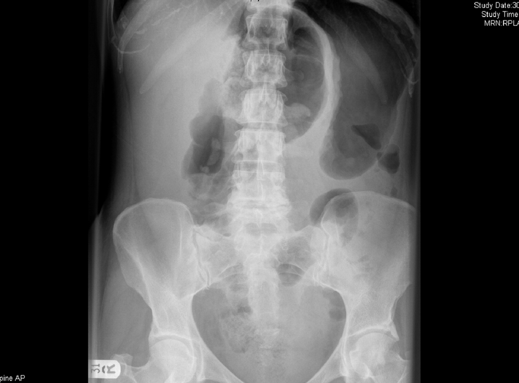
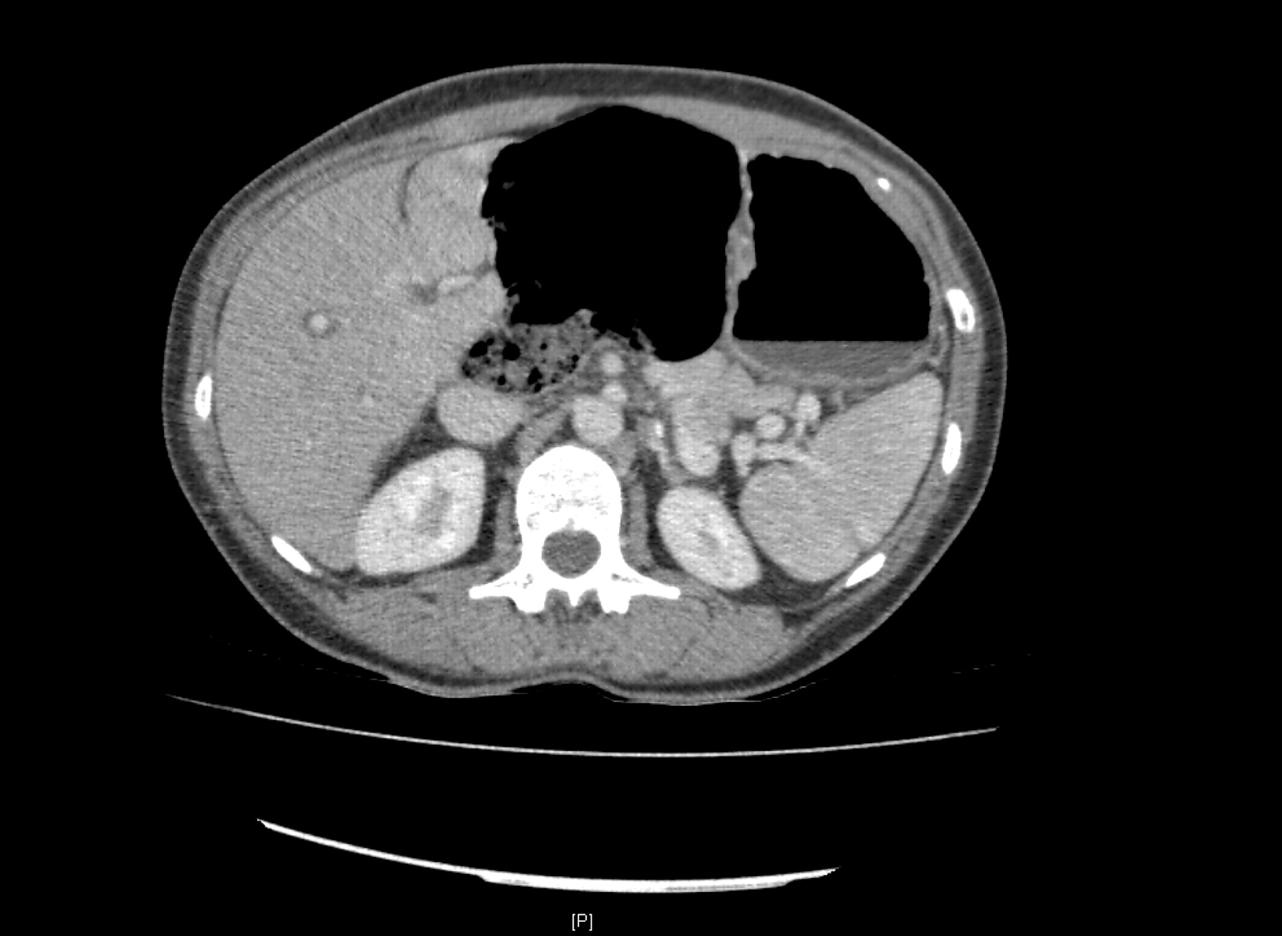
Full blood count, urea and electrolytes, liver function tests, and C reactive protein (CRP) were all within normal limits. Erect chest x ray and abdominal radiograph revealed a distended stomach pushed upwards and laterally by another distended bowel loop (fig 1). A computed tomography (CT) scan with contrast was performed subsequently which demonstrated that the caecum was herniated through the epiploic foramen causing gastric outlet obstruction (figs 2 and 3). There was also associated spiralling of mesenteric vessels to the right colon which was suggestive of caecal volvulus.

Plain radiograph of abdomen showing distended stomach pushed upwards and laterally by gas filled bowel loop.

Contrast computed tomography scan suggesting caecal volvulus causing gastric outlet obstruction.

Position of the caecum between the liver and the pancreas, suggesting possible herniation through the epiploic foramen.
TREATMENT
The patient received initial management with intravenous fluid, nasogastric tube drainage, and anti-sickness and analgesic medications. After fluid resuscitation, the patient underwent an exploratory laparotomy. A mobile caecum and ascending colon with a long, unfixed mesentery was herniated through the epiploic foramen. The caecum was twisted 180° inside the lesser sac, compressing the distal part of the stomach. The twisted right colonic mesentery was also compressing the proximal duodenum (fig 4). The hernia was reduced by milking the air and fluid contents into the transverse colon. A right hemicolectomy with ileo-transverse colonic anastomosis was performed. A portion of greater omentum was mobilised from the transverse colon and placed in the epiploic foramen to prevent further herniation.

Diagram of perioperative findings (drawn by author)—twisted and taut mesentery of ascending colon marked with an arrow.
OUTCOME AND FOLLOW-UP
The patient’s postoperative recovery was uneventful and she was discharged after 4 days.
DISCUSSION
Caecal herniation through the epiploic foramen is a rare cause of intestinal obstruction and preoperative diagnosis is reached rarely. If the caecum herniates through the epiploic foramen the “hilum” of the kidney shaped loop of bowel points towards the epiploic foramen rather than the right iliac fossa, as found in caecal volvulus.5 Interestingly, both components were present in this case and the CT scan showed the “hilum” pointing towards the epiploic foramen (fig 2). Caecal volvulus was suggested in this case by spiralling of the mesenteric blood vessels. Twisting of the mesenteric vessels (“whirl sign”) on CT scan has been described as a diagnostic sign of volvulus in literature.6
Though herniation of the caecum and ileum through the epiploic foramen has been reported, causing small bowel obstruction,3,4 this is the first reported case causing gastric outlet obstruction. Apart from herniated and distended caecal volvulus, the taut caecal mesentery across the duodenum was also responsible for obstruction. All the predisposing factors described in the literature, including an enlarged foramen of Winslow, mobile caecum and ascending colon with long mesentery, were present in this case.3 Although the exact reason for volvulus of the herniated caecum was not known in this case, the hyperperistalsis of the obstructed caecum and stomach in reverse direction might be the possible precipitating factor.
Osvaldt et al reported that delay in the treatment of internal hernia through the epiploic foramen is responsible for high mortality rates of around 36–49%.1 Obstruction, strangulation and perforation with associated metabolic and septic sequelae are the major complications of this condition.7 Clinical examination and suspicious x ray findings can lead to further investigation in the form of contrast CT scan, as in this case. An experienced radiologist could promptly reach the diagnosis in an exceptional situation like this.8
LEARNING POINTS
-
Ptotic caecum with a long mesentery might get twisted and herniated through a wide epiploic foramen, giving rise to gastric outlet obstruction without signs of small bowel obstruction.
-
If suspected clinically and on abdominal radiograph, a contrast CT scan of the abdomen can interpret the pathology effectively.
-
Early operative intervention in the form of a right hemicolectomy can save the early and late complications of the condition.
Footnotes
Competing interests: none.
Patient consent: Patient/guardian consent was obtained for publication