Article Text

Abstract
An immunocompetent male in his 70s was diagnosed with psoriatic arthritis based on dactylitis, onycholysis of the nails and scalp psoriasis. He was treated with corticosteroids, methotrexate and local corticosteroid injections without improvements in his symptoms. When tumor necrosis factor-alpha inhibitor treatment was introduced, the symptoms worsened and dactylitis of all digits and a bluish-red rash were observed on the extensor side of the left hand and arm. At this point, a skin biopsy was performed showing histopathological changes compatible with Lyme borreliosis and serum contained IgG antibodies against Borrelia burgdorferi. It was concluded that he was suffering from acrodermatitis chronica atrophicans (ACA) and Lyme dactylitis. Ten days of phenoxymethylpenicillin treatment was initiated, and after 2 weeks, the dactylitis and ACA had regressed substantially. After 6 months, both had resolved. This case emphasises the need for clinical reassessment, when treatment is not effective.
- Rheumatology
- Dermatology
- Infectious diseases
- Bone and joint infections
This is an open access article distributed in accordance with the Creative Commons Attribution Non Commercial (CC BY-NC 4.0) license, which permits others to distribute, remix, adapt, build upon this work non-commercially, and license their derivative works on different terms, provided the original work is properly cited and the use is non-commercial. See: http://creativecommons.org/licenses/by-nc/4.0/.
Statistics from Altmetric.com
Background
Dactylitis is the diffuse swelling of an entire digit typically related to an underlying inflammation or an infiltrative disorder. Psoriatic arthritis (PsA) is the most common disease that causes dactylitis due to joint, tendon and soft tissue inflammation.1 However, dactylitis can also be a clinical feature in, for example, ankylosing spondylitis, reactive arthritis, granulomatous diseases, malignancies and infections.2–5
Lyme borreliosis (LB) is a tick-borne disease caused by spirochetes within the Borrelia burgdorferi sensu lato (B.b.s.l.) complex. There are several human pathogenic genospecies within the complex, but Lyme arthritis is most often associated with B. burgdorferi sensu stricto, B. afzelii with skin lesions and B. garinii and B. bavariensis with neuroborreliosis.6 7 LB is endemic in several areas of Europe and the USA. The clinical manifestations of LB are diverse. The most common is erythema migrans (EM) at the tick bite site; however, a rash is not obligatory.8 9 Other manifestations that can occur weeks to years after the infective tick bite are neuroborreliosis, acrodermatitis chronica atrophicans (ACA), lymphocytoma cutis, arthritis, carditis, lymphadenopathy and multiple EM.10 11 Diagnosis of LB is based on characteristic clinical signs and symptoms complemented by serological confirmation of infection, once an antibody response has been mounted.6 We present a case of a patient initially presumed to be suffering from PsA, but with symptoms worsening during tumor necrosis factor-alpha (TNF-α) inhibitor treatment. A reassessment of the diagnosis showed LB with dactylitis, arthritis and ACA. The patient was successfully treated with phenoxymethylpenicillin (PcV).
Case presentation
An immunocompetent man in his 70s was seen in a rheumatological outpatient clinic at a Danish regional hospital. He was previously diagnosed with type 2 diabetes, atrial flutter, chronic obstructive lung disease and chronic myelomonocytic leukaemia; all comorbidities were well treated. One year previously, he had been diagnosed with PsA based on dactylitis of the second and third digits of the left hand, onycholysis of the nails and scalp psoriasis. The dactylitis was not painful and he had no systemic symptoms of inflammation such as fever or malaise. Treatment with corticosteroids and methotrexate did not have any effect on the dactylitis. Ultrasound-guided corticosteroid injections for tenosynovitis in the two digits (triamcinolone hexacetonide) did not have any effects on the dactylitis; hence, TNF-α inhibitor treatment(adalimumab 40 mg every 2 weeks) was initiated. At 3 months’ follow-up, the severity of the dactylitis had worsened, substantially involving all fingers of the left hand. Also, a bluish-red discolouration and induration of the skin were observed on the left upper extremity involving thenar as well as hypothenar and extending proximally and around the elbow. Underlying blood vessels related to the involved fingers were accentuated (figure 1). The skin lesions were found to be indicative of ACA, and treatment with methotrexate and TNF-α inhibitor was stopped. Since the patient had not responded as expected to the corticosteroids or disease-modifying antirheumatic drugs, the patients diagnosis was questioned. Therefore, a skin biopsy was performed showing histopathological changes compatible with LB. The patient also had anti-B.b.s.l. IgG antibodies in serum. It was concluded that the patient was not suffering from PsA but LB with manifestations of dactylitis, arthritis and ACA. Treatment with PcV 1.5 million IU three times a day for 10 days was initiated. This treatment was chosen based on recommendations from a specialist in infectious diseases. After 2 weeks, the dactylitis had regressed substantially (figure 2), and after 6 months, the dactylitis and skin lesions had vanished (figure 3). However, the patient suffered from sequalae from the left hand in terms of persistently reduced movements in both the second and third digits in both proximal and distal interphalangeal joints, despite occupational therapy. The patient did not recall having been bitten by a tick. He did recall that the first swelling of the fingers emerged weeks after gardening and removal of ivy.

Lyme borreliosis with manifestations of dactylitis, arthritis and acrodermatitis chronica atrophicans. Left hand prior to phenoxymethylpenicillin treatment.

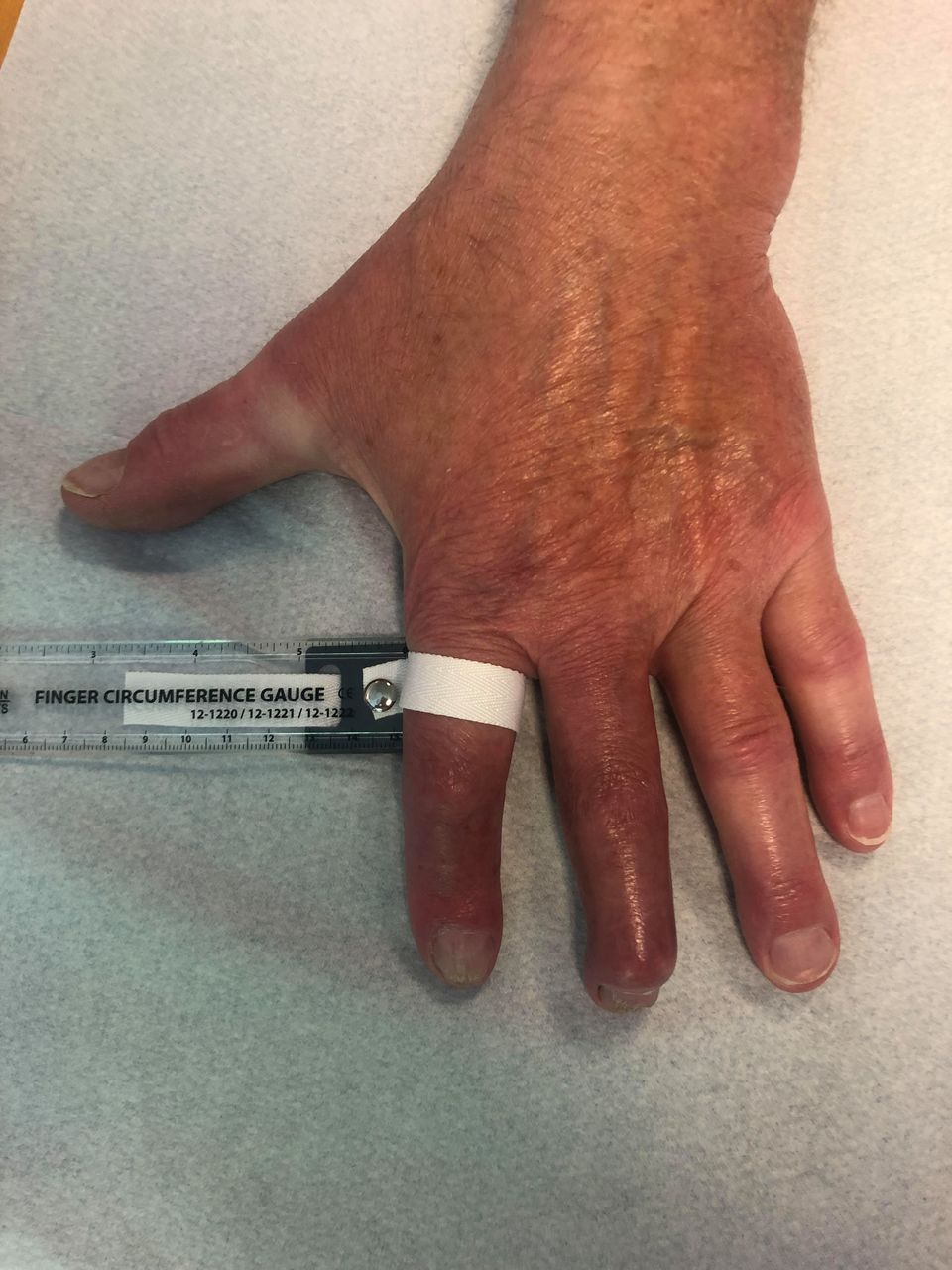
The left hand 2 weeks following phenoxymethylpenicillin treatment.

The left hand 6 months following phenoxymethylpenicillin treatment.
Investigations
Blood samples were drawn from the patient on his first visit to the Department of Rheumatology. No inflammation was present: total leucocyte count was 3.82 (ref. 3.50–8.80×) 109/L and serum C reactive protein (CRP) level was 5.8 (ref.<6.0) mg/L, serum urate level was 0.31 (ref. 0.23–0.48) mmol/L and negative findings for anticyclic citrullinated peptides, IgM rheumatoid factor and myeloma protein, and there were no antibodies against hepatitis B, hepatitis C virus or parvovirus B19. Immunoglobulins were normal. Interferon gamma release testwas also negative. During the disease course, testing for antinuclear antibodies, antineutrophil cytoplasmic antibodies, antiphospholipids, cryoglobulins and angiotensin-converting enzyme was conducted. All analyses were within the normal range. However, B.b.s.l.-specific IgG antibodies were found using the liaison Borrelia IgM and IgG CLIA assay (Diasorin, Saluggia, Italy).
X-rays of the hands and feet showed normal bone and joints. A CT scan of the thorax, abdomen and pelvis showed reactive enlarged lymph nodules in the left axil corresponding to the involved left hand. Ultrasound investigations of the fingers showed severe soft-tissue swelling, flexor tenosynovitis and synovitis of all interphalangeal joints. A skin biopsy from the left hand was performed. It was fixated using 4% formaldehyde and stained using H&E. The histopathological investigations showed deep perivascular infiltration of inflammatory cells, primarily lymphocytes, and some histiocytes and eosinophils. Also, deep and superficial plasma cells and a mild degree of spongiosis were described. The skin biopsy did not show granulomatous changes. The histopathological findings could be compatible with LB. It was not possible to perform specific PCR for B.b.s.l. as the skin biopsy had been fixated in formaldehyde.
Differential diagnosis
In this case, LB was initially misdiagnosed as PsA. Other diagnostic considerations for the causes of dactylitis were malignancy including progression in known haematological disease, granulomatous disease such as tuberculosis and sarcoidosis, lymphocytoma cutis and slow-growing infection. Idiopathic lymphoedema and discolouration of the fingers due to microthromboembolic disease were also taken into diagnostic consideration.
Treatment
When the diagnosis LB was confirmed, a specialist in infectious diseases was consulted. Treatment with PcV 1.5 million IU three times a day for 10 days was recommended. We decided to await further improvement without elongation or adjustment of the antibiotic medication as initial treatment was effective. Alongside the medical treatment, in the following 6 months, he also received occupational therapy.
Outcome and follow-up
During follow-up, inflammatory parameters remained within the normal range including CRP repeated monthly. The circumference of the digits was measured using a finger circumference gauge. Before treatment with PcV, after 2 weeks and after 6 months, the circumferences of the second digit were 11.5, 10,0 and 9.5 cm, respectively. After 6 months, tenosynovitis, arthritis, soft tissue swelling and skin rash had resolved, assessed clinically and by ultrasound. The patient was considered cured of the infection. However, he suffered from sequelae in terms of impaired flexion of the left hand’s second and third digits in the distal and proximal interphalangeal joint despite occupational therapy.
Discussion
We report a case of LB initially misdiagnosed as PsA. The symptoms were progressive dactylitis, arthritis and skin rash. There was no history of a tick bite or EM. The clinical manifestations worsened, following treatment with TNF-α inhibitor. LB was diagnosed by typical manifestation of ACA, histopathological changes in a skin biopsy, anti-B.b.s.l. IgG antibodies in serum and effect of treatment. A CT scan confirmed local lymphadenopathy.
PsA is a heterogeneous disease that can be seen with symmetrical or asymmetrical joint involvement. Dactylitis in PsA is reported in 40%–50% of patients and can be either acute with swelling, redness, and pain or chronic with swelling without pain.12 TNF-α inhibitors are effective in the treatment of dactylitis.13 In our case, dactylitis worsened when treated with an immunosuppressive TNF-α inhibitor, which led us to reassess the diagnosis.
Although dactylitis is a common feature of PsA, it may also be seen as a clinical feature in several other diseases. First, dactylitis may be seen in other rheumatic diseases such as spondyloarthritis and reactive arthritis,14–16 and in microcrystalline deposition diseases including gout and calcium hydroxyapatite with calcific tendinitis and deposition of crystals in joints and soft tissue.5 Second, dactylitis may be seen in granulomatous diseases like sarcoidosis and tuberculosis17–19 and in other slow-growing infections, for example, atypical mycobacterium infection4 and brucellosis.20 Third, in case of a more acute disease course, highly virulent pyogenic infections like Staphylococcus and Streptococcus may be considered.5 Finally, in children with dactylitis, benign osteoid osteoma,21 sickle cell disease and congenital syphilis may be considered.5 22
Clinical symptoms in LB are not always specific, and LB can look like several diseases.23 It can cause multisystem disease, particularly affecting the skin, musculoskeletal system, nervous system and heart. Lyme arthritis is usually not painful and is not accompanied by fever. It has been shown that 84% of patients have monarthritis and that the knees are affected in up to 98%.24 ACA is reported in 1%–3% of LB cases in Europe.6 ACA and arthritis are late manifestations of LB and may present months to years after the infectious tick bite.6 9
To our knowledge, this is the first case of LB describing asymmetrical dactylitis in a hand. Previously, a case report of LB dactylitis in a toe has been published by Levy et al.3 Also, diffuse hand and finger swelling has been reported as a striking feature of Lyme arthritis.25 Borrelia lymphocytoma cutis is a non-tender bluish-red nodule that may mimic dactylitis if it is located in one digit.10 However, it is typically found on the ear lobe, areola mammae or scrotum.6
The diagnosis of LB may be challenging and the diagnosis of disseminated infection is often delayed several years because of the similarity of this entity to other diseases.26 One of the reasons is that the patients do not always recall a preceding tick bite and dermatological manifestations are not obligatory. Furthermore, once a patient has developed anti-B.b.s.l. antibodies, these can persist for years.27 B.b.s.l.-specific PCR has a reasonable sensitivity in Lyme arthritis, but with a higher sensitivity in synovial samples than on synovitis fluid (range 40%–96%) but a high specificity (100%).6 In our case, it was not possible to confirm the diagnosis from PCR of the skin biopsy as the biopsy had been fixated using formaldehyde, but this could have supported the diagnosis. Culture in skin and synovial tissue/fluid is time-consuming and costly and is not recommended in routine diagnostic testing.6 The diagnosis may further be validated by the histopathological pattern in LB, including varying degrees of superficial and deep perivascular and interstitial lymphocytic infiltrates mixed with plasma cells and eosinophile. Epidermal changes such as spongiosis are seen in some cases. However, the histological features of EM and ACA are not specific.9 23
Depending on geographical variations and the clinical manifestations, different treatments regimens for LB are recommended.6 9 In Denmark, PcV for 21 days is recommended as first-line antibiotic treatment for Lyme arthritis and ACA, whereas doxycycline and ceftriaxone are alternatives to PcV.28 29 However, recommendations for treatment of LB are based on small case series, but there are no randomised controlled trials. European treatment guidelines for LB with arthritis or ACA often recommend doxycycline for 14–28 days as first-line treatment or alternatively amoxicillin or ceftriaxone.6 24 30–33 In this case, treatment with PcV for only 10 days was used with success. If treatment with PcV had not been sufficient, extension of treatment for a total of 21 days or change in treatment to doxycycline or ceftriaxone would have been considered.33
In conclusion, dactylitis is a common feature of PsA. However, dactylitis may also be seen as a clinical feature in several other diseases such as LB and should be considered both initially and if treatment response is lacking.
Patient’s perspective
The worst part of the disease was the uncertainty regarding the diagnosis. I often wondered, ‘Why can the doctors not figure out what is wrong with my fingers’. At the same time, I thought, ‘Well, I am not 25 years old anymore, but an elderly man, and I am not as vain as I used to be’. I was really happy when I was sent to the dermatologist and the skin biopsy helped to set the diagnosis, borreliosis. Already after 1 week of penicillin treatment, the swelling of my ‘sausage fingers’ improved. I have to live with a pair of semi-stiff fingers, but I think I will manage it well, even though I have to say good morning to those distorted ‘bandits’ every day.
Learning points
Dactylitis is a common feature of PsA. However, dactylitis may also be seen as a clinical feature in several other diseases such as Lyme borreliosis (LB).
LB may occur although a tick bite or a history thereof or an initial erythematous rash is not recalled by the patient.
When suspecting an infection, attention to the correct procedure for culture and PCR analysis is important.
Other differential diagnoses must be considered when expected treatment response is lacking. It is crucial to re-evaluate the medical history, objective examinations and laboratory tests. In this case, the conclusive clue was hidden in the skin biopsy.
Ethics statements
Patient consent for publication
Acknowledgments
We thank the patient for participating with his personal experience and letting us write the case report. We would like to thank Mette Ramsing, Department of Pathology, Sygehus Lillebælt, for performing the pathological investigations and Axel Møller, Department of Infectious Diseases, Sygehus Lillebælt, Denmark, for helping to set the right diagnosis. We thank Henrik Thorman for performing the skin biopsy.
References
Footnotes
Contributors CMA treated the patient in the outpatient clinic and provided pictures and the patient's perspective. AS, CMA and NSA wrote and reviewed the case report.
Funding The authors have not declared a specific grant for this research from any funding agency in the public, commercial or not-for-profit sectors.
Case reports provide a valuable learning resource for the scientific community and can indicate areas of interest for future research. They should not be used in isolation to guide treatment choices or public health policy.
Competing interests None declared.
Provenance and peer review Not commissioned; externally peer reviewed.