Article Text

Abstract
The authors report two cases of unusually large deposits on their therapeutic bandage contact lens (BCL) following uneventful surgery for congenital ptosis. The first case presented at 6 weeks with decreased vision, large jelly-bump deposits over the contact lens and sterile corneal infiltrates. The infiltrates rapidly resolved with restoration of vision following contact lens removal and topical antibiotics. The second case presented 2 weeks after surgery with visual loss and similar deposits but with no corneal involvement. Following replacement of BCL and topical lubricants, her vision improved to 20/20. Studies on the role of BCL in ptosis surgery are scarce with literature supporting its use for ocular surface protection and minimising postoperative discomfort. The authors hypothesise impaired blink mechanism as the accelerating factor for this unusual occurrence in the early postoperative period and recommend frequent replacement of the contact lens and a closer follow-up in all these cases.
- Ophthalmology
- Medical management
Statistics from Altmetric.com
Description
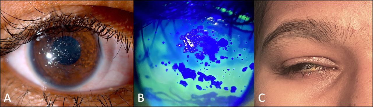
We describe two patients with jelly-bump deposits on the therapeutic contact lens used for corneal protection postptosis surgery. The first was a woman in her 20s who underwent uneventful left levator resection surgery for moderate simple congenital ptosis. Dry eye and lagophthalmos were ruled out preoperatively. Senofilcon-A therapeutic bandage contact lens (BCL) (Johnson & Johnson) was placed at the end of surgery for protection against corneal exposure. Carboxymethyl cellulose 1% eye-drops 2 hourly, and ofloxacin 0.3% ointment over the skin were prescribed. She was lost to follow-up. Six weeks later, she reported with decreased vision. Her best-corrected visual acuity (BCVA) had decreased to finger counting at 1 m. Examination revealed a congested eye, a retained contact lens with large jelly-bump deposits, and mucoid film over the BCL (figure 1A, B). She had 3 mm lagophthalmos with minimal corneal exposure (figure 1C). The underlying cornea revealed multiple superficial infiltrates. Microbiological evaluation of BCL and corneal scrapings did not reveal any micro-organisms. Intensive broad-spectrum topical antibiotics were started, and the corneal lesions decreased in intensity in 3 days, improving the BCVA to 20/40, with residual corneal infiltrates. Subsequently, she followed up locally and her vision improved to 20/20.

Case 1 (A) and (B) showing circumciliary congestion with large, jelly-bump deposits in the left eye. (C) Postoperative lagophthalmos with upto one-third cornea exposed.
The second case was a teenaged female who underwent repeat levator resection surgery for right residual simple congenital ptosis. Senofilcon-A BCL (Johnson & Johnson) was placed at the end of the surgery. There was good Bell’s phenomenon and no dry eye preoperatively. Two weeks later, BCVA dropped from 20/20 to 20/400. Examination revealed large jelly-bump deposits over the BCL and 3 mm lagophthalmos (figure 2A–C). The BCL was replaced with resultant improvement in the visual acuity to 20/20.
Case 2 (A) Jelly-bump deposits over the bandage contact lens involving the visual axis, highlighted as (B) negative uptake of fluorescein stain. (C) Postoperative lagophthalmos with reduced blink rate.
First described in 1970s, ‘jelly-bump’ deposits, also known as mulberry spots or lens calculi, are discrete or confluent, elevated lesions on contact lenses; comprising cholesterol and waxy esters.1 2 Senofilcon-A CLs used for extended wear have higher oxygen transmissibility however they also have higher propensity for jelly-bump deposition compared with conventional soft CL. Factors known to cause rapid deposition on the lens include concurrent dry eye, meibomian gland dysfunction, and systemic disease.3 In an oculoplastic setting, therapeutic CLs have been commonly used for cornea protection and enhance patient comfort following ptosis repair.4 The current cases developed CL deposits despite the use of lubricants and in the absence of dry eye, leading to sterile corneal infiltrates in the first case. The authors attribute postoperative impaired blink reflex and insufficient lid closure as the possible mechanism for the accelerated and exuberant deposits. Reduced blink reflex impedes the movement of the lid over the ocular surface permitting accumulation of tear-mucous material. This promotes microbial colonisation which potentially can lead to microbial keratitis.
Following ptosis surgery, the improvement in the ocular surface parameters is gradual.5 The early postoperative period is crucial, necessitating a contact lens in select cases. This is the first report illustrating the postoperative development of jelly-bumps over therapeutic CLs following ptosis surgery. This finding may perhaps be overlooked due to lack of knowledge about this entity and its implications.
Learning points
Impaired blink reflex following ptosis surgery may accelerate deposition of jelly bumps over silicone hydrogel contact lenses (CLs).
The clinician should be mindful of this rare occurrence while placing CL for ptosis surgery as it can lead to development of keratitis and its complications.
The authors recommend closer follow-up in the early postoperative period and early replacement of the CL to avoid this complication.
Ethics statements
Patient consent for publication
Footnotes
Contributors All persons designated as authors qualify for authorship, and all those who qualify are listed. Each author has participated sufficiently in the work to take public responsibility for appropriate portions of the content. AA collected the data, conceptualised and drafted the manuscript. SIM edited the manuscript. MNN, NB and SIM finalised the article. NB and MNN were involved in the management of the patients and contributed to the interpretation of the results.
Funding This study was funded by Hyderabad Eye Research Foundation (LEC-BHR-R-08-22-927).
Case reports provide a valuable learning resource for the scientific community and can indicate areas of interest for future research. They should not be used in isolation to guide treatment choices or public health policy.
Competing interests None declared.
Provenance and peer review Not commissioned; externally peer reviewed.