Article Text

Statistics from Altmetric.com
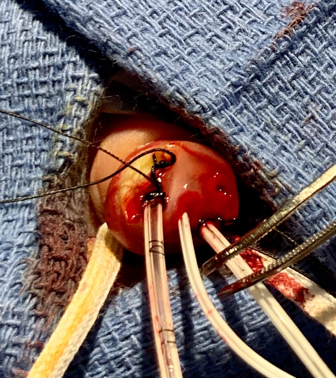
Description
A full-term neonate was born to a gravida 2 para 1 mother at 38 weeks gestational age via spontaneous vaginal delivery. The pregnancy was uncomplicated. An anatomy scan done at 22 weeks gestational age was normal except for the presence of an isolated left ventricular echogenic intracardiac focus. The amniotic fluid was meconium stained and infant had poor respiratory efforts at birth requiring CPAP (continuous positive airway pressure) in the delivery room. The Apgar scores were 5, 6 and 8 at 1 min, 5 min and 10 min, respectively. Infant was admitted to the NICU (neonatal intensive care unit) for high oxygen requirement on CPAP and received in and out surfactant × 1 dose. Umbilical lines were placed on admission to the NICU in view of high oxygen requirement and potential for developing persistent pulmonary hypertension. Infant was discovered to have four-vessel umbilical cord (two arteries and two veins) as shown in figure 1. An echocardiogram done on day of life 1 showed left aortic arch with mild discrete isthmus hypoplasia with no coarctation of aorta and a tiny patent foramen ovale. Follow-up echocardiogram done 1 month later as outpatient was normal except for the presence of patent foramen ovale. An abdominal ultrasound revealed no anomalies. Infant was weaned off from CPAP by day 3 of life and discharged home on day of life 6.

Four-vessel umbilical cord in a newborn showing two arteries and two veins.
Four-vessel umbilical cord with persistence of right umbilical vein is a rare finding with incidence of 1: 526.1 2 Four-vessel umbilical cord may be associated with multiple congenital malformations.3 It may be diagnosed prenatally on ultrasound and postnatal diagnosis is usually made during the placement of umbilical lines.
Learning points
A thorough examination of newborns should include counting of the umbilical cord vessels.
Infants found to have four-vessel umbilical cord should get a detailed evaluation, including abdominal ultrasound and cardiac echo to rule out associated anomalies.
Ethics statements
Patient consent for publication
Footnotes
Contributors RA wrote the initial draft of the manuscript and performed literature search. PA helped in literature search and editing of the initial version. CT-D as supervising neonatologist envisioned the idea, performed final editing and obtained patient permission and consent.
Funding The authors have not declared a specific grant for this research from any funding agency in the public, commercial or not-for-profit sectors.
Case reports provide a valuable learning resource for the scientific community and can indicate areas of interest for future research. They should not be used in isolation to guide treatment choices or public health policy.
Competing interests None declared.
Provenance and peer review Not commissioned; externally peer reviewed.