Article Text

Statistics from Altmetric.com
Description
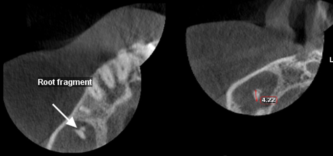
A middle- aged patient was referred for Cone beam CT (CBCT) of right maxilla. The patient had undergone extraction of a right second premolar and the dentist was suspecting root fracture. CBCT of right maxilla was done to aid in retrieving the location of root tip. Image acquisition was done using Planmeca ProMax 3Ds machine (Planmeca, Helsinki, Finland) with standard operating specifications. Planmeca Romexis software V.3.01 R was used for viewing and analysing the images. CBCT slices were analysed in axial, coronal and sagittal sections. Sagittal section showed extraction socket in tooth 15 region. Sagittal section showed a well-defined radiopacity within the right maxillary sinus with perforation of floor of right maxillary sinus (figure 1). Axial section confirmed presence of root fragment within the antrum suggestive of root fragment, measuring about 4.22 mm (figure 2). Mucosal thickening was evident in the right maxillary sinus suggestive of sinusitis (figures 1 and 2). Radiographically, a diagnosis of oroantral communication (OAC) was given. The patient was referred for surgical evaluation for treating OAC and Otolaryngologists for further evaluation of sinus pathology. OAC is an unpleasant complicated situation encountered by dental practitioners where an abnormal communication develops between oral cavity and the maxillary sinus.1 It is always better to diagnose OAC as early as possible and should be closed within 24 hours to prevent any sinus infection and inappropriate healing of the socket.2 A limitation of two-dimensional imaging; however, is the superimposition of anatomical structures.3 However, CBCT overcomes this limitation and can be used to identify OAC and also to determine the status of the soft tissue in the maxillary sinus and to identify sinus pathology, which proved to be of high diagnostic value in the reported case.

Sagittal section shows perforation of the floor of the maxillary sinus.

Axial section shows evidence of root fragment and its measurement.
Learning points
The most common cause for oroantral communication (OAC) is the extraction of maxillary posterior teeth. This may be due to the proximity of the roots to the maxillary sinus floor, which is also exacerbated by the presence of thin floor of the maxillary sinus.
Imaging modalities should always be used in conjunction to diagnose OAC. However, OAC is diagnosed clinically.
Soft tissue changes in maxillary sinus due to disruption of maxillary sinus floor should be taken care with priority.
Ethics statements
Patient consent for publication
Footnotes
Contributors PMD has contributed to the conception or design of the work; and interpretation of data for the work; and final approval of the version to be published. VN has contributed to drafting the work or revising it critically for important intellectual content; and final approval of the version to be published.
Funding The authors have not declared a specific grant for this research from any funding agency in the public, commercial or not-for-profit sectors.
Case reports provide a valuable learning resource for the scientific community and can indicate areas of interest for future research. They should not be used in isolation to guide treatment choices or public health policy.
Competing interests None declared.
Provenance and peer review Not commissioned; externally peer reviewed.