Article Text

Statistics from Altmetric.com
Description
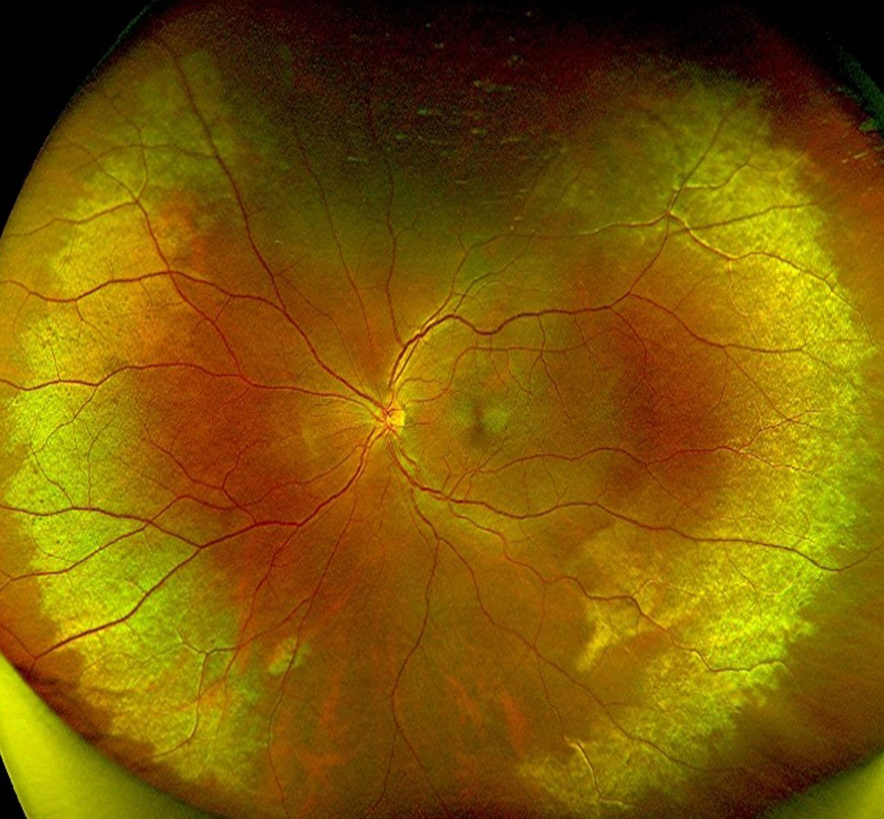
The diagnosis of commotio retinae is clinical and relies on funduscopic examination. The classic finding is retinal whitening or opacification that clinically resolves within 4– 7 days.1 Disruption of the photoreceptor outer segments is the primary underlying pathology with sparing of the blood vessels.2 When the macula is involved, it is known as Berlin’s oedema, with a ‘pseudo cherry red spot’.3 Though a majority of patients recover in 1–4 weeks with no permanent deficits, those with severe commotio may have residual retinal pigment epithelium (RPE) changes, RPE atrophy or hyperpigmentation.4 5 A healthy man around 40 years of age presented to us after sustaining an injury to the left eye with a tennis ball. Anterior segment showed diffuse conjunctival congestion, iris sphincter tears and open angles on gonioscopy. The visual acuity and intraocular pressure were normal. Fundus examination showed an interesting distribution of commotio retina in the mid-periphery concentric to the disc but sparing the posterior pole (figure 1). The equatorial expansion after the severe blunt trauma with a uniformly shaped tennis ball could have resulted in this pattern. Commotio retinae usually presents with a localised or diffuse pattern and seen in the inferior and medial retinal periphery as these are the most vulnerable areas. The present case highlights an unusual pattern of commotion retinae concentric around the disc and should prompt a closer follow-up for other sequelae in view of the severity of the injury.

Fundus picture of the left eye showing concentric retinal whitening suggestive of commotio retinae.
Learning points
Commotio retinae is a consequence of blunt ocular trauma and the presentation can indicate the severity of the impact.
While commotio retinae self resolves in a majority, a close follow-up is mandatory to monitor for other sequelae of trauma.
Ethics statements
Patient consent for publication
Footnotes
Contributors SS collated the data, CJ and SGK wrote the manuscript.
Funding The authors have not declared a specific grant for this research from any funding agency in the public, commercial or not-for-profit sectors.
Case reports provide a valuable learning resource for the scientific community and can indicate areas of interest for future research. They should not be used in isolation to guide treatment choices or public health policy.
Competing interests None declared.
Provenance and peer review Not commissioned; externally peer reviewed.