Article Text

Statistics from Altmetric.com
Description
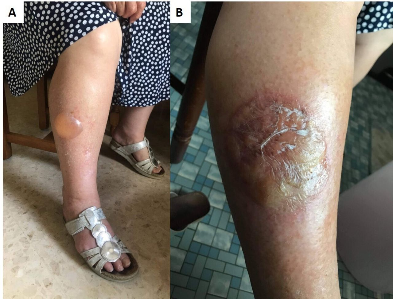
A woman in her 80s with type 2 diabetes diagnosed 10 years prior, hypertension, low-grade carotid artery stenosis, stage 2 chronic kidney disease, hypothyroidism and severe obesity arrived at the consultation with a 7-cm-wide tense-roof bulla on the dorsum of the right foot that had developed 2 days prior and prevented her from wearing closed shoes (figure 1). The temperature was 37.1°C, the heart rate was 74 beats/min and the blood pressure was 138/96 mm Hg. There was no pruritus, no surrounding inflammation, no Nikolski’s sign, no Asboe-Hansen sign, a normal monofilament test result, no mucosal lesion and perceived tibial pulses. Daily treatment included acetylsalicylic acid 75 mg, hydrochlorothiazide 25 mg, irbesartan 300 mg, gliclazide 60 mg, levothyroxine sodium 50 g and metformin 850 mg, plus dulaglutide 0.75 µg as one subcutaneous injection per week. No cognitive impairment existed; there had been no trauma or recent new-drug intake; the glycated haemoglobin had been below 7.5% for more than a year; and the follow-up eye exam revealed no signs of diabetic retinopathy. A blood test showed haemoglobin at 11 g/dL, leucocytes at 6.7 g/L (neutrophils: 4.49 g/L; eosinophils: 0.14 g/L), glucose at 7.48 mmol/L, glycated haemoglobin at 7.5%, creatinine at 76 µmol/L, serum albumin at 32 g/L, C reactive protein less than 10 mg/L and normal liver enzymes. The right-foot blister ruptured spontaneously, leaving an erythematous wound that healed in 1 month with the triweekly application of hydrocellular foam dressings. A second blister formed on the right leg (figure 2) and immediately ruptured. A biopsy of the post-bullous erythema revealed angiodermatitis with papillary and reticular dermal angiogenesis, neovessel endothelial turgidity and lymphocytic perivascular inflammation. Direct immunofluorescence studies were negative. Cultures were also negative. Bullosis diabeticorum (BD) was diagnosed based on these findings and the clinical history.

(A) Sizeable blister of the dorsum of the right foot. (B) Post-bullous erythema at day 7. (C) Right-foot post-bullous lesion at day 15.

(A) Bullous lesion on the anterolateral face of the right leg. (B) Post-bullous erythema at day 15.
Other blisters formed on the lower limbs in the following months, causing pain, daily discomfort and footwear issues; then they ceased. At the 1-year consultation, no new blisters had developed in the past 6 months, the glycated haemoglobin level was 7.3%, which is optimal because it was below 7.5%,1 and there was still no diabetic retinopathy or peripheral neuropathy.
BD is an infrequent, non-inflammatory, diabetes-related skin complication.2 The differential diagnosis includes bullous pemphigoid, friction bullae, oedema-induced bullae, bullous fixed drug reaction and epidermolysis bullosa acquisita.2–4 The annual incidence is expected to be 0.16% in patients with diabetes mellitus.5 Though the prognosis is favourable, BD may impair quality of life, particularly if the blister is large.6 7 To avoid infectious or haemorrhagic complications and achieve a quick healing time, strict local care is required.2 3 Although the aetiology appears to be multifactorial, abnormal calcium and carbohydrate metabolism and microangiopathy are believed to be the main culprits.2–4 7
Better glycaemic control and screening for microangiopathy complications are recommended because BD is more likely in patients who have long-standing uncontrolled diabetes and peripheral neuropathy.4 5 8 Our clinical observation suggests that disabling BD may develop in the elderly despite optimal glycaemic control and few to no microangiopathy complications.
Learning points
Bullosis diabeticorum is characterised by recurrent, spontaneous, non-inflammatory bullae in the setting of long-term, uncontrolled diabetes. Although the prognosis is good, the risks of infection, haemorrhage and delayed healing are increased in the case of a large blister.
Lesions on the lower limbs can make it difficult to wear shoes, cause discomfort and necessitate extensive care, resulting in a significant reduction in the elderly’s quality of life.
Bullosis diabeticorum should be investigated for microangiopathic complications of diabetes, although it may occur in their absence.
Ethics statements
Patient consent for publication
Footnotes
Contributors Both authors cared for the patient and collected the data. RH did literature work, and wrote the drafted the manuscript. Both authors revised the manuscript and supervised every process.
Funding The authors have not declared a specific grant for this research from any funding agency in the public, commercial or not-for-profit sectors.
Case reports provide a valuable learning resource for the scientific community and can indicate areas of interest for future research. They should not be used in isolation to guide treatment choices or public health policy.
Competing interests None declared.
Provenance and peer review Not commissioned; externally peer reviewed.