Article Text

Statistics from Altmetric.com
Description
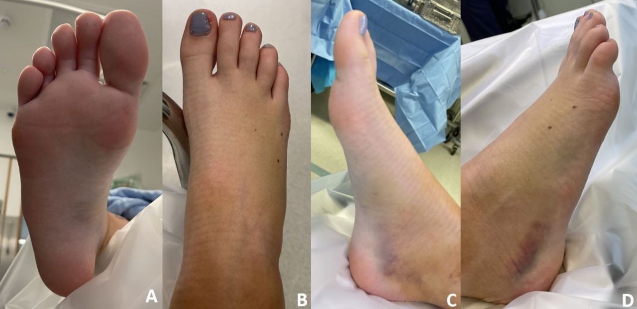
A woman in her late teens presented to the trauma clinic with an isolated fracture–dislocation of the right navicular bone after falling from a scooter. She had no significant background medical history and was otherwise fit and healthy, with no history of smoking or diabetes mellitus. On examination, she exhibited noticeable swelling, point tenderness on the dorsomedial surface and ecchymosis on the plantar, medial and lateral aspects of the midfoot and hindfoot, without any neurovascular deficit (figure 1).

Preoperative clinical photograph showing (A) plantar, (B) dorsal, (C) medial and (D) lateral views of the foot. Note the ecchymosis on the plantar, medial and lateral aspects.
CT scan of the right foot showed a heavily comminuted fracture of the navicular with multiple displaced bony fragments (figure 2). No other fracture was identified. There was an otherwise normal alignment of the ankle and midfoot, and the Lisfranc ligament was intact. She subsequently underwent open reduction and internal fixation with two dissolvable magnesium screws and a bridging plate (figures 3 and 4). Initially, she was weight-bearing as tolerated in a walking boot for 6 weeks, and then subsequently weaned from the boot. She underwent removal of the bridging plate 2 months later, and her postoperative course was uncomplicated with an excellent functional outcome (figure 5).

Preoperative (A) axial, (B) sagittal and (C) coronal CT slices demonstrating the comminuted navicular fracture.

Intraoperative clinical photograph showing a Hintermann distractor allowing for anatomic reduction of the comminuted fracture–dislocation.

Intraoperative fluoroscopic imaging showing anatomic reduction with bridging plate and two dissolvable magnesium screws in good position.

Postoperative plain flims after removal of bridging plate with two dissolvable magnesium screws in situ.
The navicular plays an important role in the biomechanics of the midfoot. It articulates distally with the cuneiform bones, proximally with the talus and laterally with the cuboid bone. There are thick ligamentous attachments both plantar and dorsal, attaching the navicular to surrounding structures.1 These extensive attachments make an isolated fracture–dislocation to the navicular bone very rare. The navicular bone has a precarious blood supply and is at risk of developing avascular necrosis, necessitating careful consideration in the reduction and fixation of these fractures.2 Navicular fractures are responsible for approximately 5% of all foot fractures.3
Isolated talonavicular fracture–dislocations are rare injuries, and as far as the authors are aware, only a handful of case reports are available in the literature. The majority of cases are managed by closed reduction and fixation with Kirschner wires.4 Careful consideration should be given to the management of these fractures due to the incidence of associated injuries as well as the important role that the navicular bone plays in the biomechanics of the midfoot. The use of magnesium alloy screws in conjunction with a temporary bridging plate proved to be effective in this case.
Patient’s perspective
My accident occurred when I decided to come down a considerably steep hill on a scooter. As I was coming down the hill, I didn’t realise the speed I had picked up until I was seconds away from hitting a kerb and being thrown from the scooter. As a result of the speed and impact of the fall, I broke my foot. Immediately after the fall, there was no pain until I tried to stand on my right foot. Two days later, my foot doubled in size and I was brought for an X-ray. Over the 2 days, I was able to wiggle my toes and wasn’t in excruciating pain and therefore did not anticipate the damage I had done to my foot.
Learning points
The navicular plays an important role in the biomechanics of the midfoot, articulating distally with the cuneiform bones, proximally with the talus and laterally with the cuboid bone.
There are thick ligaments that attach the navicular to surrounding structures, and the sheer strength of these extensive attachments make an isolated fracture–dislocation to the navicular bone very rare.
Careful consideration should be given to the management of these fractures due to the incidence of associated injuries as well as the important role that the navicular bone plays in the biomechanics of the midfoot.
Ethics statements
Patient consent for publication
Footnotes
Contributors RH, MNR and OH wrote the manuscript. OH and TB treated the patient.
Funding The authors have not declared a specific grant for this research from any funding agency in the public, commercial or not-for-profit sectors.
Case reports provide a valuable learning resource for the scientific community and can indicate areas of interest for future research. They should not be used in isolation to guide treatment choices or public health policy.
Competing interests None declared.
Provenance and peer review Not commissioned; externally peer reviewed.