Article Text

Statistics from Altmetric.com
Description
A 62-year-old man underwent coronary CT angiography for evaluation of suspected atherosclerotic coronary artery disease (CAD). While no obstructive CAD was noted, a unique left atrial anomaly was incidentally observed. A smooth hypodense fibromuscular band-like structure was noted within the left atrium, connecting the anteromedial and posterior walls of the left atrium and partially covering the right inferior pulmonary vein (RIPV) ostium (figures 1 and 2).
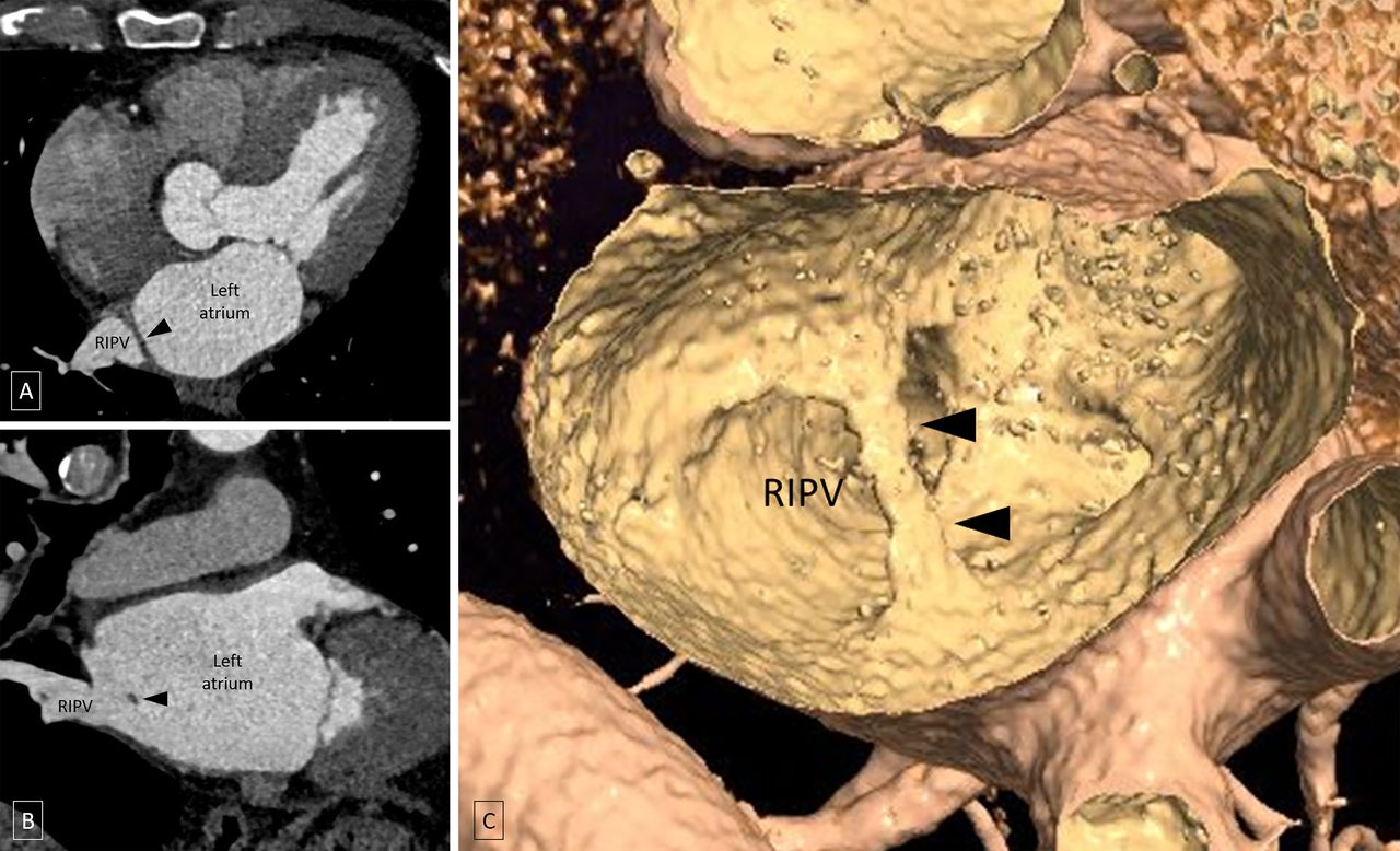
Oblique axial (A) and oblique coronal (B) images of CT angiography and virtual dissection image (C) show a smooth band-like structure (black arrowheads) within the left atrium, connecting its anteromedial and posterior walls, and partially covering the right inferior pulmonary vein (RIPV) ostium.
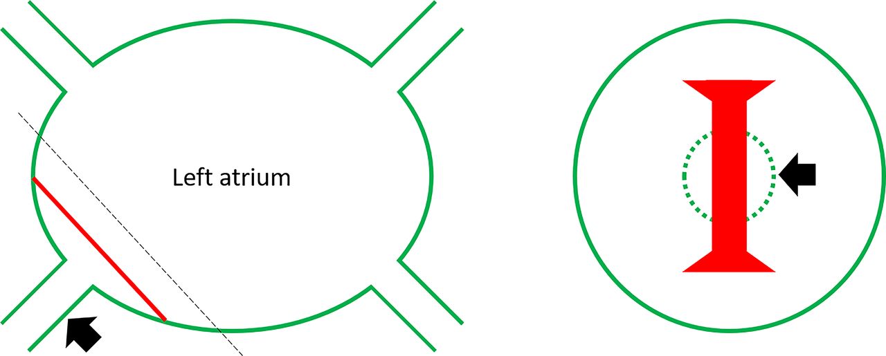
Schematic shows the smooth left atrial band (red line) connecting the anteromedial and posterior wall of the left atrium in relation to the right inferior pulmonary vein (RIPV) (thick black arrow) ostium. An en-face view from the plane of the dotted line shows the band partially covering the RIPV ostium (dotted green circle). (The figure is illustrated by N.N.P.)
The left atrial band is a rare congenital anomaly with an incidence of 2% in autopsy studies.1 In most of the described cases, the band is seen connecting the left atrial aspect of the fossa ovalis with various areas of the left atrial endocardium. Various histopathological studies have demonstrated that these bands are composed of fibromuscular tissue. Although a benign entity, it has potential clinical implications. It is associated with the presence of Chiari network, premature atrial complexes, patent foramen ovale, mitral valve prolapse and regurgitation and episodes of cardioembolism.1 2 The presence of the left atrial band, especially in relation to the pulmonary venous ostia as seen in the current case, can pose technical challenges during radiofrequency catheter ablation in the setting of atrial fibrillation.3 The present case highlights the role of CT angiography in the complete anatomical depiction of the left atrium and associated anomalies.
Learning points
Left atrial band is a rare congenital anomaly.
It is associated with the presence of Chiari network, premature atrial complexes, patent foramen ovale, mitral valve prolapse and regurgitation and episodes of cardioembolism.
CT angiography plays an important role in the complete anatomical depiction of the left atrium and associated anomalies.
Ethics statements
Patient consent for publication
Footnotes
Contributors MV: prepared the manuscript. NNP: prepared the manuscript and images including the line diagram. SK: edited and proofread the manuscript. RY: edited and proofread the manuscript.
Funding The authors have not declared a specific grant for this research from any funding agency in the public, commercial or not-for-profit sectors.
Case reports provide a valuable learning resource for the scientific community and can indicate areas of interest for future research. They should not be used in isolation to guide treatment choices or public health policy.
Competing interests None declared.
Provenance and peer review Not commissioned; externally peer reviewed.