Article Text

Statistics from Altmetric.com
Description
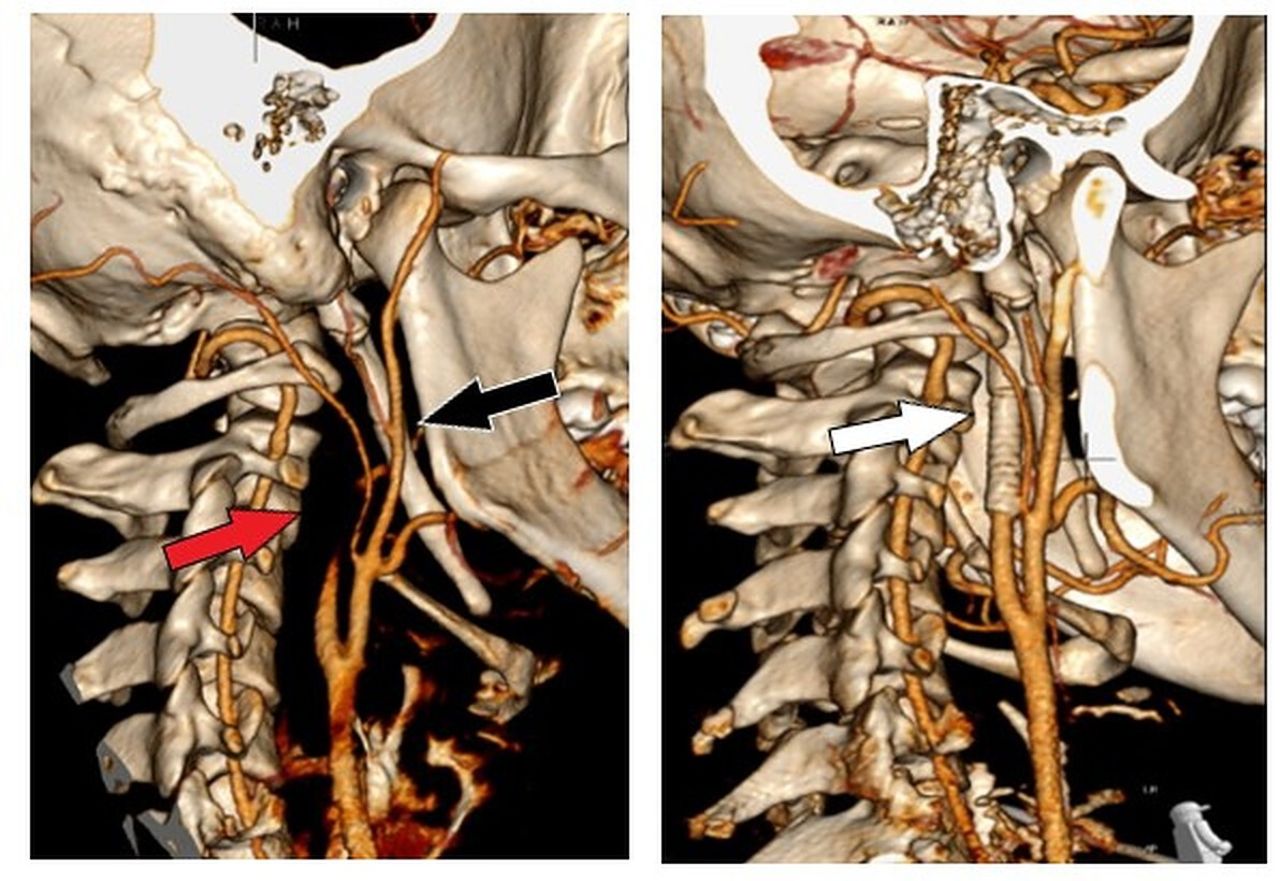
A young student was resistance training at the gym when he developed a headache and collapsed. On waking, there was left-sided facial weakness, visual extinction and sensory loss. Investigations revealed a right internal carotid artery dissection (ICAD) and M1 thrombus. Intravenous thrombolysis was given, but he deteriorated (National Institutes of Health Stroke Scale, 14 to 20). He was referred to interventional radiology and successfully treated with mechanical clot retrieval and ICAD stenting (figure 1). A right frontal lobe infarct was evident on CT (figure 2). Dual antiplatelets were prescribed for 3 months followed by lifelong single antiplatelet. He had no stroke risk factors or medical history. On reviewing his CT images, bilateral elongated styloid processes (ESP) were identified; 7.4 cm for the right and 7.0 cm for the left (normal less than 3 cm) (figure 1). The patient declined stylectomy, but he made a full recovery and the only problem at 3 years is low mood.

Left: reconstructed CT image demonstrating absence of flow in right internal carotid artery (red arrow) and right-sided elongated styloid process which has two pseudoarticulations (black arrow). Right: Postprocedure reconstructed image demonstrating stent adjacent to styloid process (white arrow).

Head CT demonstrating evolving infarction of right frontal lobe (white arrow).
An elongated styloid process, which can include a calcified stylohyoid ligament, does not usually cause symptoms.1 Rarely, some develop pain due to contact with neurovascular structures in what is known as Eagle syndrome after the otolaryngologist describing it in 1937.2 Our patient never had symptoms of Eagle syndrome. ICAD due to ESP is rare, but well described.3 4 Some authors have identified ESP length or ESP angulation and carotid artery proximity as being important risk factors for ICAD, but others have demonstrated that these factors have no relation.5–7 Triggering events like exercise and neck massage are commonly seen, but most are spontaneous.3 4 The initial treatment of ICAD due to ESP resembles that of ICAD and stroke, with many authors opting for medical management with or without early endovascular intervention.3 4 Like our patient, many with ICAD due to ESP have early deterioration despite medical management, so they should be monitored with a low threshold for referral to interventional radiology or otolaryngology. Definitive treatment of Eagle syndrome is resection, but the role of ESP resection for ICAD is unclear.8 Late complications can occur in patients without ESP resection, namely recurrent stroke, carotid stent fracture, persistent ICA stenosis with dissection flap, stent displacement and pseudoaneurysm formation, so stylectomy or delayed imaging should be considered.9–12
Learning points
Dissection is a common cause of ischaemic stroke in young patients.
In patients with spontaneous or low energy dissection, review images for an elongated styloid process.
Consider monitoring in these patients as early and late deterioration is reported.
Ethics statements
Patient consent for publication
Footnotes
Contributors MT composed the manuscript. LI directed the project. AS and DJW were responsible for interpreting imaging. All authors were involved in the design and review of the manuscript and the literature.
Funding The authors have not declared a specific grant for this research from any funding agency in the public, commercial or not-for-profit sectors.
Competing interests None declared.
Provenance and peer review Not commissioned; externally peer-reviewed.