Article Text

Statistics from Altmetric.com
Description
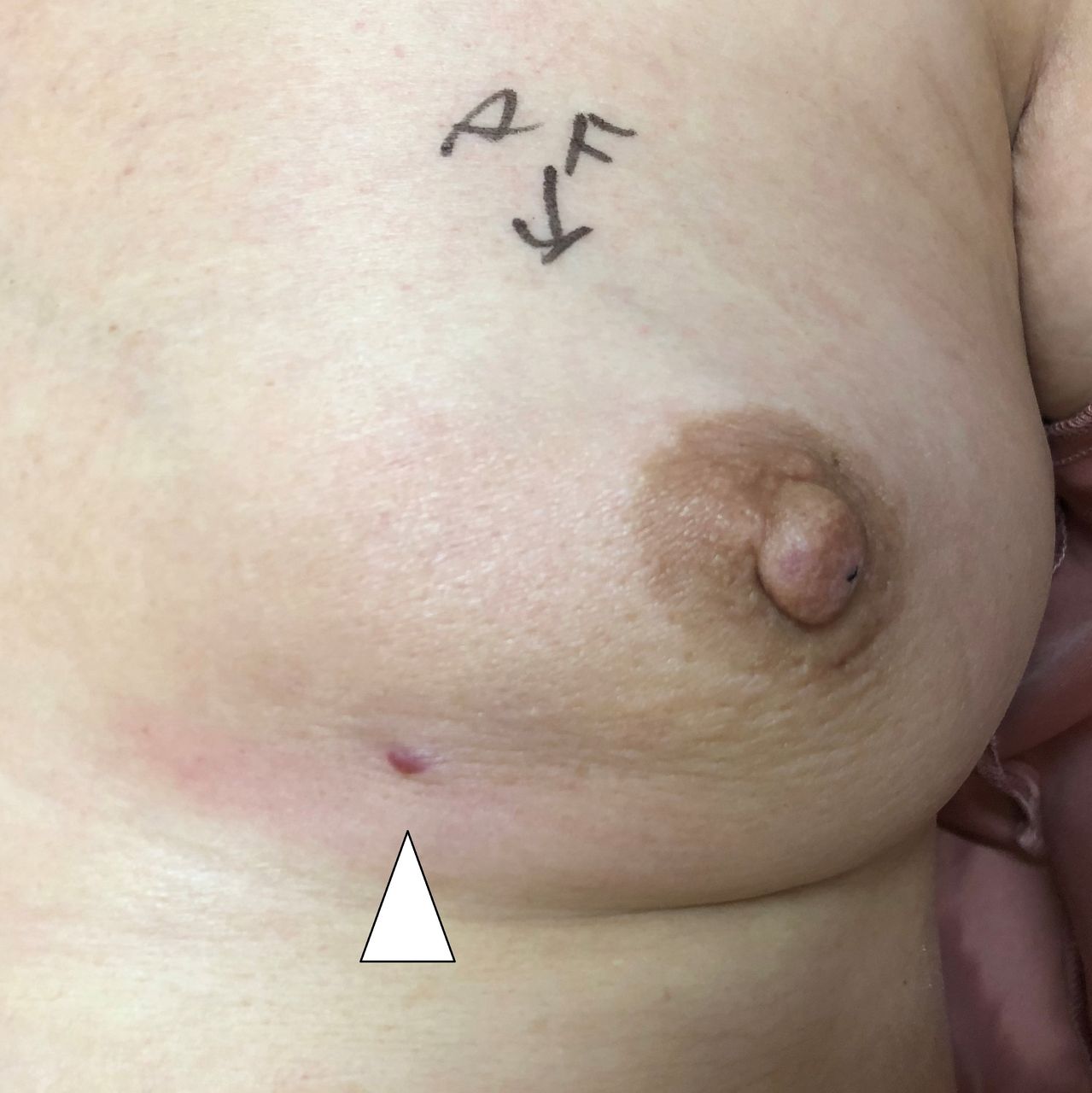
A 71-year-old woman with an abnormality detected during the mammography screening consulted a physician. The physician palpated a mass measuring approximately 2 cm in the left lower outer quadrant, with no abnormal skin findings. Ultrasonography revealed an 18 mm hypoechoic mass with irregular margins in this region. A 12-gauge needle vacuum-assisted biopsy (VAB) (Celero; Hologic, Marlborough, MA) was performed. A puncture was made in the lower inner quadrant, approximately 5 cm inward from the tumour. She was diagnosed with breast cancer and presented to our hospital for treatment. On palpation (33 days postbiopsy), a new reddish nodule was observed at the puncture site with induration underneath it (figure 1). Ultrasonography (35 days) revealed a cord-like hypoechoic region from the tumour to the skin nodule, which was the biopsy route (figure 2A). Additionally, hypervascularised tissue was found around this area on colour Doppler. Imaging using 18F-fluorodeoxyglucose positron emission tomography/CT (47 days) (figure 2B) and contrast-enhanced MRI (48 days) (figure 2C) also revealed a cord-like lesion from the tumour to the skin nodule suggesting that the cancer had progressed through the biopsy route. Left mastectomy and axillary lymph node dissection were performed; the skin nodule and biopsy route were completely resected (70 days). Based on the pathological results, the patient was diagnosed with pT4bN1M0 stage IIIB invasive breast cancer. Immunohistochemical staining revealed positive expressions of oestrogen and progesterone receptors and a human epidermal growth factor receptor-2 score of 2+. Consistent with preoperative imaging, skin infiltrative lesions, consisting primarily of lymphatic vessel tumour emboli were found between the tumour and the skin nodule. Furthermore, the formation of cancerous lesions with dermis infiltration were observed in the skin nodule. The total infiltration diameter extended to 84 mm. The cause was seeding associated with tumour progression that was mainly composed of lymphatic vessel tumour emboli in the biopsy route. In recent years, the treatment of cancer has become increasingly complex; moreover, it is important to collect and preserve an adequate amount of cancer tissue prior to commencing the treatment. In this case, since the patient was not at high risk of bleeding, we used VAB, which is effective in collecting a sufficient amount of tissue and is considered to be highly effective in diagnosis, instead of a core needle biopsy (CNB). VAB may not have a high frequency of seeding compared with CNB, since VAB requires less punctures and produces suction pressure.1 2 Additionally, it has been suggested that most cancer cells displaced by a needle biopsy do not survive.3 However, VAB, which uses a larger needle than CNB, may carry a higher risk for seeding due to the ‘suckling’ effect on the tumour and spread through the needle tract. Predicting the risk of seeding on imaging before biopsy is difficult, CNB may be safer and more recommendable in the first biopsy to avoid this rare complication. Conversely, during VAB, it is crucial to puncture from a site such that the entire biopsy route resection could be possible.

On palpation (33 days postbiopsy), a new reddish nodule noted on the skin at the puncture site (white arrowhead).

Ultrasonography revealing a cord-like hypoechoic region from the tumour to the skin nodule along the biopsy route (A) (arrow heads). 18F-fluorodeoxyglucose (FDG) positron emission tomography/CT showing significant FDG uptake at the site of the cord-like lesion (B) (arrow heads). Contrast-enhanced MRI also showing an enhanced cord-like lesion in the coronal section (C) (arrow heads).
Learning points
Tumour seeding rarely occurs in breast needle biopsies.
This is a notable case, in which seeding in the biopsy route was observed on preoperative imaging.
Performing the puncture from a site where the entire biopsy route can be resected is important.
Ethics statements
Footnotes
Contributors Conception and design of study: AF and HY. Acquisition of data: AF and TH. Analysis and/or interpretation of data: AF and TH. Drafting the manuscript: AF. Revising the manuscript critically for important intellectual content: TH and TS. Approval of the version of the manuscript to be published: AF, HY, TH and TS.
Funding The authors have not declared a specific grant for this research from any funding agency in the public, commercial or not-for-profit sectors.
Competing interests TS reports personal fees from ASKA Pharmaceutical,Astra Zeneca K.K.,Eisai, Ono Pharmaceutical,Taiho Pharmaceutical,Takeda Pharmaceutical,Chugai Pharmaceutical, Eli Lilly Japan K.K.,Novartis Pharma K.K.,Pfizer, MiRTeL,Meiji Seika Pharma Nippon Kayaku, grants from Eisai.,Kyowa Hakko Kirin,Taiho Pharmaceutical,Chugai Pharmaceutical,Nippon Kayaku, outside the submitted work.
Provenance and peer review Not commissioned; externally peer reviewed.