Article Text

Statistics from Altmetric.com
Description
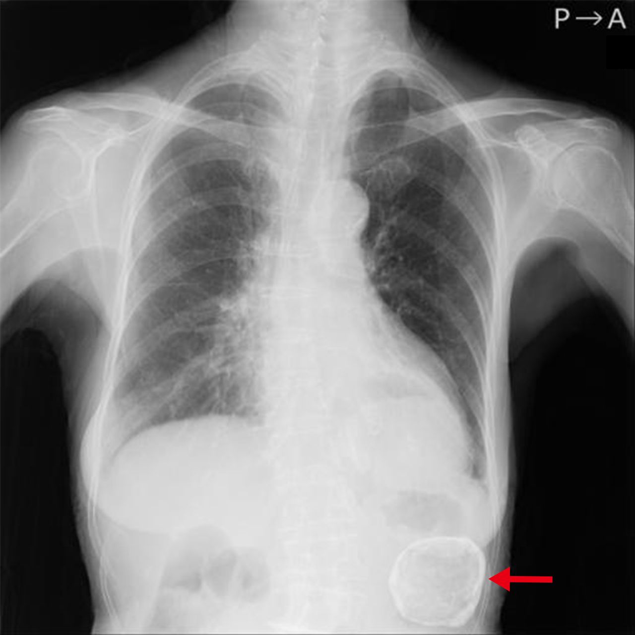
A 93-year-old female with Alzheimer’s dementia underwent a routine plain chest radiography before admission to a nursing home, which incidentally showed an abdominal mass (figure 1). The patient had been seen twice a month by our clinic’s visiting physicians and had been doing well with no particular symptoms. Laboratory examinations showed no remarkable findings. Abdominal CT revealed a splenic mass with a diameter of 58 mm with calcification at the margins, indicating the diagnosis of epidermoid cyst (figure 2). We practised shared decision making with the patient and her family, and it was decided that no further detailed examination or treatment would be performed on the splenic tumour.

Plain chest radiography showing the abdominal mass with calcified margins (red arrow).

CT showing the splenic mass with wall calcification (yellow arrows) (A, B, axial views; C, coronal view; D, sagittal view).
Incidental abnormal lesions are defined as abnormalities unrelated to the patient’s presenting illness. In the context of the spleen, these lesions mean that the patient has no history of malignancy and no symptoms, such as fever, weight loss and upper abdominal discomfort. Unexpected splenic lesions are frequently identified on CT in daily clinical practice. Although most lesions are benign, clinicians should be alert to warning findings as follows: the presence of solid, contrast-enhancing components and ill-defined lesion borders.1 These findings suggest the possibility of potentially more relevant diseases, such as malignancy, abscess and sarcoidosis. Alternatively, splenic incidentaloma detected on chest X-ray is rare. To the best of our knowledge, this is the first case of incidentally identified splenic masses on plain chest radiography.
Splenic epidermoid cysts are uncommon congenital benign lesions. They are a form of non-parasitic primary cysts. They are composed of cystic lesions lined by keratinising and nonkeratinising epithelia, which may be squamous or cuboidal. Symptoms of epidermoid splenic cysts are usually nonspecific. The most common symptoms are a left upper abdominal mass, followed by left upper abdominal pain.2 Complications from epidermoid splenic cysts, such as bleeding, infection and rupture, are rare. On imaging examinations, epidermoid of the spleen has a solitary, well-defined cystic lesion, occasionally with plaque-like wall calcification. Surgical therapy is often recommended for symptomatic patients and for cysts larger than 5 cm. The goal of the surgical treatment is to avoid complications and prevent recurrence.3
Routine chest radiography obtained on admission has been a common practice in many hospitals. In addition to that, especially in Japan, routine chest X-rays are often performed before admission to nursing homes mainly to rule out tuberculosis (since Japan is still a tuberculosis middle-burden country).4 Clinicians should pay attention to the potential existence of abnormal findings outside the lung fields when reading routine plain chest radiographic images.
Learning points
Incidentaloma on plain chest radiography is a rare clinical presentation of splenic masses.
Splenic epidermoid cysts are uncommon congenital benign lesions and rarely lead to complications, including bleeding, infection and rupture.
Clinicians should be aware of the possibility of abnormal findings in areas other than the lung fields when reading routine plain chest radiographic images.
Footnotes
Contributors HF acquired data and drafted the manuscript. MH reviewed and supervised the manuscript.
Funding The authors have not declared a specific grant for this research from any funding agency in the public, commercial or not-for-profit sectors.
Competing interests None declared.
Provenance and peer review Not commissioned; externally peer reviewed.