Article Text

Abstract
Urachal cancer is a rare and aggressive cancer that often presents in advanced stages. Given the rarity of this malignancy, medical case studies provide one of the few sources of literature available through which clinicians can guide medical management. Surgery is widely considered to be the mainstay of therapy when disease is localised and surgically resectable, therefore most current case studies on urachal cancer focus on surgical management, occasionally with adjuvant chemotherapy. However, few case studies discuss chemotherapy alone in the treatment of metastatic disease. Most studies indicate a median overall survival between 12 and 24 months for metastatic urachal adenocarcinoma. Bone marrow metastasis of solid tumours, when considered alone, portends a poor prognosis. The patient in this case study represents a rare case of stage IV urachal adenocarcinoma metastatic to the bone marrow without progression of disease after 6 months of treatment.
- urological surgery
- urological cancer
- gynaecological cancer
- chemotherapy
This is an open access article distributed in accordance with the Creative Commons Attribution Non Commercial (CC BY-NC 4.0) license, which permits others to distribute, remix, adapt, build upon this work non-commercially, and license their derivative works on different terms, provided the original work is properly cited and the use is non-commercial. See: http://creativecommons.org/licenses/by-nc/4.0/.
Statistics from Altmetric.com
Background
The urachus is a fibrous, tubular vestigial remnant derived from the involution of two contiguous embryonic structures, one of which is the allantois, a derivative of the yolk sac, and the other being the cloaca, a cephalic extension of the urogenital sinus which is a precursor to the fetal bladder.1–3 The urachus usually involutes after the third trimester and is obliterated by fibrous proliferation thus remaining as the medial umbilical ligament, connecting the apex of the bladder dome to the umbilicus in the midline, without any remaining physiological function.2 4 5
Urachal cancer is a rare, aggressive and often clinically silent disease due to its extravesical and extraperitoneal location.1 6 7 It accounts for less than 0.5% of bladder cancers and only 0.01% of all adult malignancies.6 8 9 Urachal cancer typically involves the dome of the bladder and is most often histologically characterised as adenocarcinoma in 80%–90% of cases, although the normal urachus is primarily lined by transitional epithelium.2 4 10 One proposed theory is that this occurs due to columnar metaplasia of the urachal mucosa while a competing theory suggests that this is due to malignant transformation of enteric epithelial rests within the urachal remnant left behind from the cloaca during embryological development.2 6 11 Histological subtype of urachal cancer most often reveals mucinous type in 50%–75% of cases, although 15%–25% of cases are enteric type, resembling colorectal adenocarcinomas and 6%–7% are signet ring cell type.10 12 13 The median age at the time of diagnosis is between 50 and 58 years old based on previous studies, which is much younger than non-urachal adenocarcinomas whose median age is around 69 years old.10 14 Urachal cancer also displays male predilection, with males representing between 60% and 68% of all cases.7 8
Patients with urachal cancer often present with locally advanced or metastatic disease, most commonly metastatic to the lungs, liver, peritoneum, lymph nodes, brain and/or bone.6 8 12 13 Given the aggressive nature of urachal cancer, early diagnosis is paramount as there is significant divergence in both treatment and prognosis depending on stage. A retrospective study performed by Mayo Clinic in Rochester, Minnesota evaluated 49 patients with urachal cancer from 1950 to 2003 and found the median overall survival for stage I/II urachal cancer to be 10.8 years vs 1.3 years for stage IV urachal cancer.8
Case presentation
A 52-year-old man with a medical history of hypertension presented to an outpatient office with shortness of breath with exertion for about 4 weeks. He also endorsed 12 pound intentional weight loss over the previous 5 months. This patient denied any haematuria, haematochezia, melena, night sweats, cough or haemoptysis. He was a non-smoker, reported infrequent alcohol use and reported no drug use. The patient’s mother had Hodgkin lymphoma and small-cell lung cancer, his brother had testicular cancer and his aunt had ovarian cancer. Outpatient laboratory studies were performed which revealed pancytopenia. The patient was subsequently sent to the hospital for further evaluation.
Investigations
CT scan of the chest, abdomen and pelvis with IV and PO contrast was performed and revealed a mixed density soft tissue mass measuring 7.2 cm×2.8 cm×3.2 cm arising from the anterior superior dome of the bladder in the midline extending toward the umbilicus along the course of the urachal remnant with punctate calcifications but without visible extension into the lumen of the bladder (figure 1). The CT scan also revealed multiple lung nodules and numerous hepatic hypodensities scattered throughout the liver both of which were concerning for metastases. There was no radiographic evidence of bone metastasis.
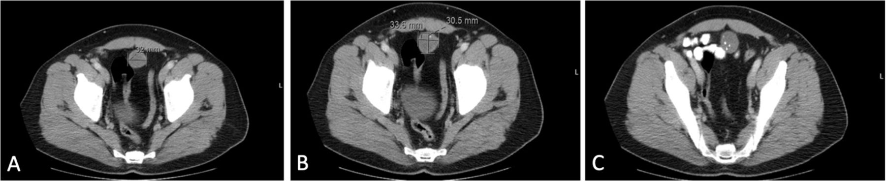
CT scan of the chest, abdomen and pelvis with IV and PO contrast revealed a mixed density soft tissue mass inseparable and arising from the anterior superior dome of the bladder extending cephalad in the midline toward the umbilicus along the course of the urachal remnant with punctate internal calcifications and no visible extension into the lumen of the bladder.
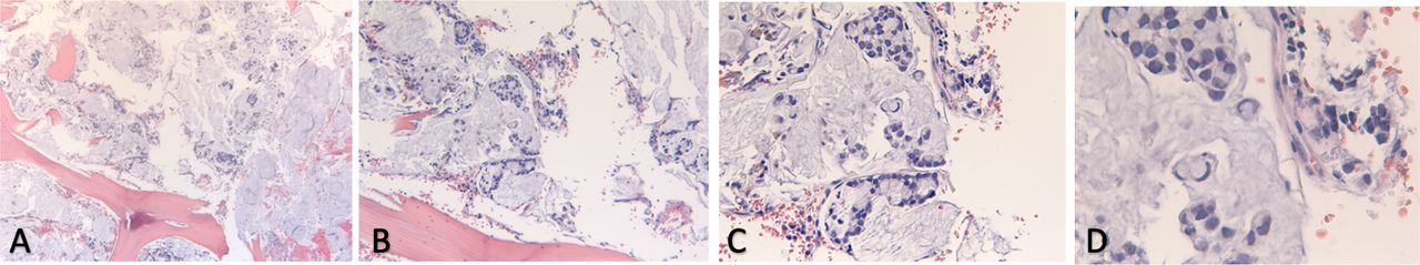
Given the patient’s pancytopenia and clinical presentation, bone marrow aspirate and biopsy were performed which revealed 90%–95% of the marrow space largely replaced by mucin within which were glandular structures composed of malignant cells including frequent signet-ring forms (figure 2). Immunohistochemical staining was positive for CK20, CDX2 and P504S and negative for CK7, TTF1 and CK903. HER2 staining was also negative. Based on pathology review and radiographic findings, this patient was diagnosed with stage IV urachal mucinous adenocarcinoma metastatic to the bone marrow with possible metastases to the liver and lung as well.
Bone marrow aspirate and biopsy revealed 90%–95% of the marrow space replaced by large lakes of mucin within which there were floating small round glandular structures composed of malignant cells with mucinous cytoplasmic vacuoles. The tumour cells included frequent signet ring forms. The little remaining bone marrow was hypercellular with maturing myeloid and erythroid precursors, likely representing erythroid hyperplasia.
Treatment
This patient was ultimately treated with a gastrointestinal chemotherapy regimen commonly known as FOLFOX-6 (Folinic acid, fluorouracil and oxaliplatin) every 14 days. Oxaliplatin was stopped after 12 cycles due to significant peripheral neuropathy. The patient was subsequently continued on maintenance therapy with 5-fluorouracil with the plan to continue fluorouracil-based treatment until progression of disease.
Outcome and follow-up
Restaging CT scan of the chest, abdomen and pelvis around 6 months after starting the aforementioned treatment regimen revealed a stable urachal mass without significant change in size, stable pulmonary nodules and slightly increased size of the hypodensities throughout the liver. His blood counts also improved with haemoglobin around 100 g/L and improved dyspnoea on exertion.
Discussion
There is no validated staging system for urachal cancer; however, the two most commonly used are the Sheldon staging system proposed in 1984 and the Mayo staging system proposed by Ashley et al. (table 1).5 7 13 15
Sheldon vs Mayo staging systems for urachal cancer
Surgery is the mainstay of therapy if localised and surgically resectable, although one-third of patients are unresectable at the time of presentation. Urachal tumours are not very radiosensitive, therefore radiotherapy is infrequently used.7 If localised and surgically resectable, a partial cystectomy with en-bloc urachectomy and umbilectomy, involving resection of the medial umbilical ligament from the bladder dome up to, and including, the umbilicus, with negative surgical margins is widely considered to be the recommended surgical approach.8 16 17 Margin-negative, en bloc resection is believed to have a significant impact on survival with several studies suggesting an increased risk of relapse when this procedure is not performed.11
Unfortunately, patients with urachal cancer often present with locally advanced or metastatic disease. However, there are limited reports of urachal cancer metastatic to the bone marrow. Bone marrow metastasis is rare, in and of itself, and patients often have a poor prognosis due to rapid disease progression and poor response to treatment.18 The most common origins of bone marrow metastasis include lung, breast, stomach and prostate.19 Chandra et al performed a retrospective study of 1419 cases that underwent bone marrow evaluation from 2006 to 2009 due to various diseases and found only 25 cases of bone marrow metastasis from solid tumours, constituting just 1.76% of all cases.20 Additionally, Anner et al evaluated 3620 bone marrow aspirates from 2877 patients with solid tumours and found 263 cases of bone marrow metastasis making up 9.1% of all cases.21 Kucukzeybek et al also performed a retrospective evaluation of a total of 3345 bone marrow biopsies and found only 58 patients with bone marrow metastasis of solid tumours with a median overall survival of just 28 days after bone marrow metastasis was discovered.19 Similarly, Zhou et al reviewed 30 patients with bone marrow metastasis and found a median overall survival time of 3 months with a median overall survival time of 9 months in the systemic therapy group.18 Taylor et al described one of the few reported cases of urachal adenocarcinoma with evidence of isolated thoracic vertebral metastasis and bone marrow involvement on biopsy; however, this patient underwent vertebral corpectomy and received no systemic chemotherapy.22
Patients with urachal adenocarcinoma metastatic to the bone marrow are not surgical candidates and chemotherapy remains the primary treatment modality in these patients. However, the role of chemotherapy in urachal cancer is not well established and there are currently no evidence-based guidelines regarding neoadjuvant or adjuvant chemotherapy for the treatment of urachal cancer.7 12 However, there is evidence to suggest that urachal cancer is less responsive to cisplatin-based chemotherapy when compared with urothelial cancer.6 Szarvas et al conducted a comprehensive meta-analysis of over 1000 cases of urachal cancer and found a higher radiographic response rate in the 5-fluorouracil-based chemotherapy regimens when compared with the cisplatin-based chemotherapy regimens (44% vs 9%, p=0.043) with the combination of both 5-fluorouracil and cisplatin chemotherapy demonstrating the lowest progression rate (14%) with a radiographic response rate of 43%.13 16 Histology, immunohistochemical staining and clinical presentation of urachal cancer classically resembles colorectal cancer and is often treated similarly.11 Given this resemblance, there have been several reports of metastatic urachal cancer responding to FOLFOX chemotherapy.7 12 23 24
The patient described in this case report demonstrates a rare case of urachal adenocarcinoma with bone marrow metastasis. Given the generally poor prognosis of both urachal cancer and bone marrow metastasis, when evaluated separately, one would surmise that prognosis would be similarly poor for a patient with both. Additionally, this patient presented with presumed metastases to the lungs and liver. The patient in this case report was classified as stage IVB based on the Sheldon staging system and stage IV based on the Mayo staging system discussed previously. Given the diffuse metastases, this patient was not considered to be a surgical candidate. Therefore, he was treated with chemotherapy (FOLFOX-6). This case report demonstrates a patient with urachal adenocarcinoma with bone marrow metastasis treated with fluorouracil-based chemotherapy without significant progression of disease after 6 months of treatment.
Learning points
Given the rarity of urachal cancer, one of the challenges is that medical case studies provide one of the few sources of literature through which clinicians have to guide medical management.
This was a rare case demonstrating that urachal adenocarcinoma can metastasise to the bone marrow which may present as pancytopenia.
This case demonstrates use of 5-fluorouracil based chemotherapy in a patient with urachal adenocarcinoma with bone marrow metastasis.
References
Footnotes
Contributors The author would like to thank Dr Brian Byrne, MD, for his contributions with analysis and interpretation of data and review of this case report.
Funding The authors have not declared a specific grant for this research from any funding agency in the public, commercial or not-for-profit sectors.
Competing interests None declared.
Provenance and peer review Not commissioned; externally peer reviewed.