Article Text

Statistics from Altmetric.com
Description
A 45-year-old man presented with a 2-year history of an enlarging left forearm mass. He has noted enlargement of his left forearm with associated pain with movement. The pain is worst with flexion on the lateral aspect of his forearm. The pain does not overtly limit his activities of daily living and improves with non-steroidal anti-inflammatory drugs. He denies numbness, tingling, weakness or unexpected weight loss. Patient recalled suffering an injury to his left forearm during a soccer game in Ghana at the age of 15 years. He was seen by a nurse who placed his forearm in a cast. No radiography images were obtained, and he had no further follow-up. He has never had issues with his forearm until the last few years. On physical examination, the patient has a notable deformity of the left forearm (figure 1). There was a palpable mass in the midulnar shaft without tenderness, erythema or skin break. His elbow and wrist range of motions were preserved with mild pain with supination, pronation and elbow flexion. He had normal strength and was neurovascularly intact. Plain radiography demonstrated a chronic and hypertrophic non-union ulnar diaphysis fracture with mild deformity of the radial diaphysis related to the healed fracture (figure 2). There was also moderate to severe osteoarthrosis of the elbow.

Left midforearm deformity (arrows) at the ulnar aspect (A and B).

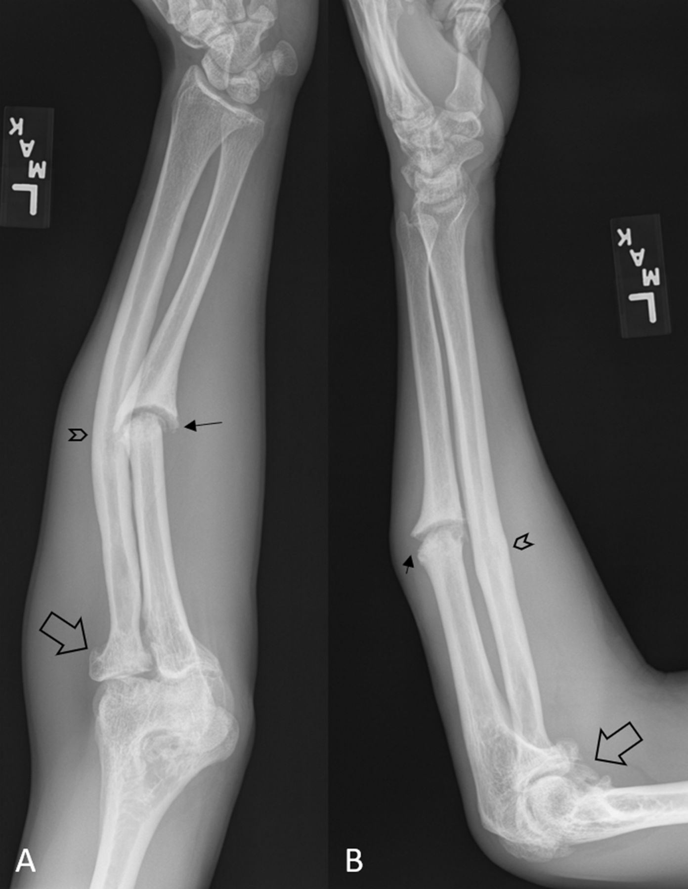
Plain anterior-posterior (A) and lateral (B) radiographs of left forearm demonstrating chronic, hypertrophic non-union (pseudarthrosis) ulnar diaphysis fracture (arrows) with deformity of radial diaphysis consistent with a healed remote fracture (arrowheads). Moderate to severe elbow osteoarthrosis is also present (open arrows).
Paediatric and adolescent forearm diaphyseal fractures present a treatment challenge.1–3 Historically, both non-surgical and surgical treatments have been performed.1 3–5 The mainstay of the management is close clinical and radiographic follow-ups. Non-unions may occur in 2%–10% of all forearm fractures and can lead to dysfunction of the upper limb and hand.1 4 Compared with children, adolescents and adults are more likely to develop malunions and non-unions.3 6 Other potential risk factors for non-unions are open fractures, inadequate stability (eg, suboptimal immobilisation or surgical fixations), associated infections and insufficient metabolic (eg, callus formation) response (eg, smoking and diabetes).1 6 Non-union can be further categorised into hypertrophic and atrophic types. Hypertrophic non-union is characterised by adequate callus formation without adequate stability.6 However, atrophic non-unions occur due to the lack of callus formation usually due to inadequate vascularity or metabolic aetiologies.6 Despite non-union, in some cases, patients may maintain reasonable function for years.1 6 7 The primary goal of treatment is to restore the functional anatomy between the radius and ulna through maintaining their respective lengths and thus obtain optimum function.1 Prolonged radial and ulnar non-union may cause significant disability due to osteoarthrosis and decreased range of motion of adjacent joints.2 Patients with malunions and non-unions should be referred to an orthopaedic surgeon for further evaluation and treatment.1 2 4 6 Our consultant orthopaedic surgeon recommended an elective ulnar open reduction and internal fixation with bone graft.6 8
Learning points
Forearm injuries require a comprehensive clinical evaluation that should include radiographic images in most cases, particularly with any deformity or loss of function.
Regardless of the treatment strategy, forearm fractures need close clinical and radiographic follow-ups.
Patients with radius or ulna non-unions should be referred to orthopaedic surgery.
Ethics statements
Patient consent for publication
Footnotes
Contributors All authors participated in patient’s care, literature review, and manuscript preparation and approval.
Funding The authors have not declared a specific grant for this research from any funding agency in the public, commercial or not-for-profit sectors.
Competing interests None declared.
Provenance and peer review Not commissioned; externally peer reviewed.