Article Text

Statistics from Altmetric.com
Description
A 66-year-old African American man with metastatic prostate cancer presented to clinic with a pruritic rash in his chest of 1 week duration. He was started on androgen-antagonist therapy with enzalutamide a month ago. Medical history was significant for chronic obstructive pulmonary disease, hypertension, post-traumatic stress disorder and hypothyroidism. His chronic medications included albuterol, tiotropium bromide, amlodipine, prochlorperazine, trazodone and levothyroxine. The lesions started as pruritic, diffusely erythematous macules with purpuric centres in the anterior and posterior thorax. Enzalutamide therapy was immediately stopped. Five days after discontinuing treatment, the patient noted progression of the rash to his entire thorax, abdomen and all four extremities in a symmetric distribution, with initial skin sloughing in his face. He denied having any intercurrent illness or influenza-like symptoms. Shortly after he was diagnosed with Stevens-Johnson syndrome/toxic epidermal necrolysis (SJS/TEN) overlap syndrome, the patient was transferred to our burn unit. Within a few hours, the patient had extensive detachment of skin involving 25% of body surface area (BSA), primarily involving superior thorax and back (figure 1). Scattered coalescing areas of skin sloughing were present in the abdomen and upper extremities. There was extension of the macular rash with vesicles and bullae formation throughout the trunk and extremities. Haemorrhagic erosions were present in the glands of the penis and the oral mucosa (figure 2). Nikolsky sign was positive. SCORTEN (TEN-specific severity-of-illness score) scoring for this patient was 3 (age≥40, presence of active/evolving malignancy and affected BSA ≥10%), corresponding with a 35% mortality rate.1 His condition quickly deteriorated needing ventilator support. The patient was managed with intravenous fluids, proper wound care and cyclosporine 4 mg/kg/day as adjunctive therapy.2 Hospital course was complicated by septic shock, ventilator-associated pneumonia, Clostridioides difficile infection and acute kidney injury needing haemodialysis. He needed multiple interventions for a perforated bladder secondary to continuous irrigation for refractory bleeding from his cancerous prostate. This resulted in an open non-healing abdominal wound. His skin lesions improved over time but unfortunately his overall clinical condition did not, and family decided to withdraw care 2 months after initial hospitalisation. The incidence of SJS/TEN is about 2–7 cases/million people per year, but mortality can be as high as 30%–40%.3 Both incidence and mortality of SJS/TEN are much higher in patients with active malignancy compared with the general population.4
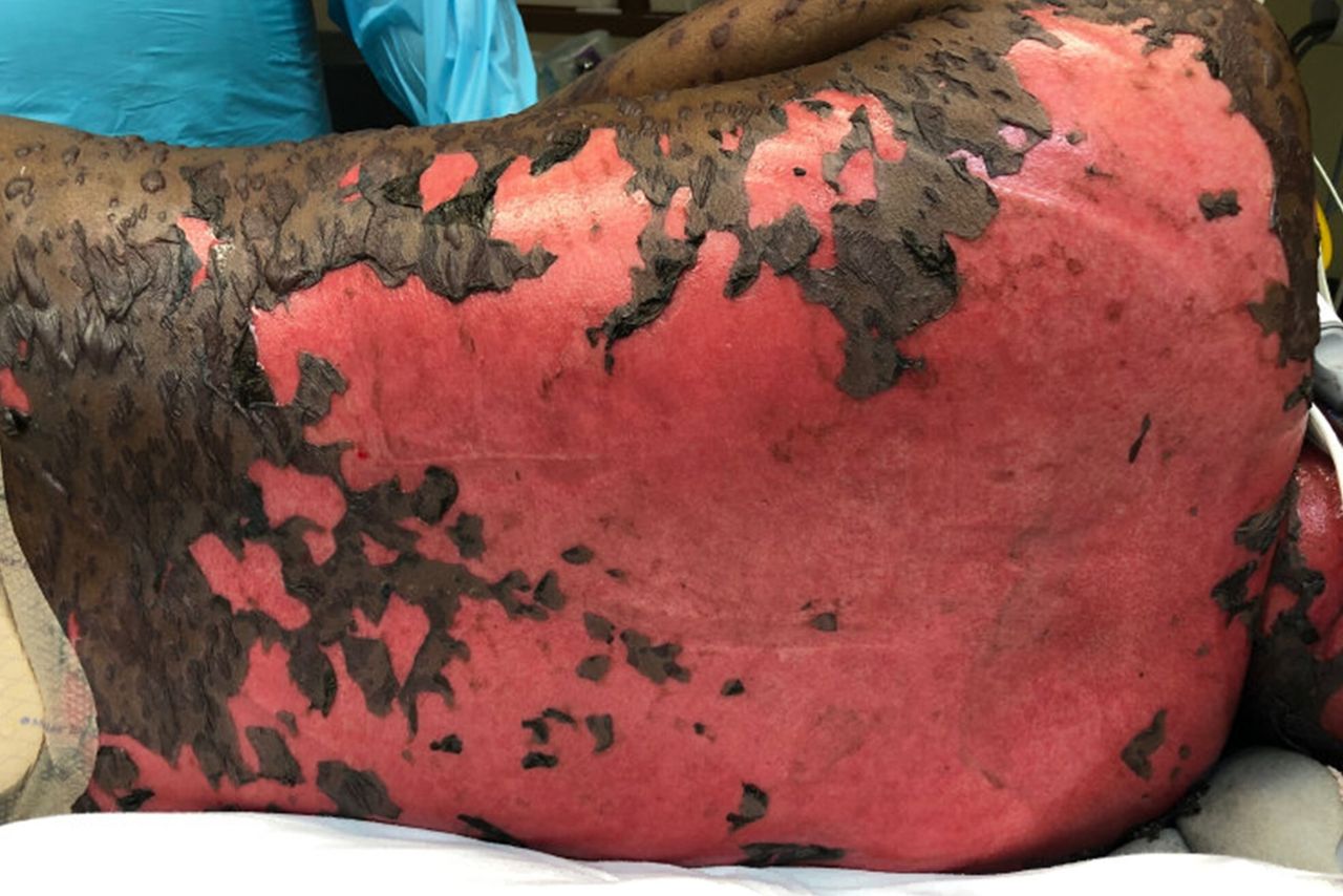
View of the patient’s back with near-complete desquamation and detachment of skin.

Haemorrhagic erosions were noted in the patient’s face and near oral mucosal surfaces. The presence of skin detachment noted in the upper chest.
Learning points
Stevens-Johnson syndrome/toxic epidermal necrolysis are characterised by epidermal necrosis and sloughing of mucous membranes and skin. They are triggered by drugs in 80%–95% cases. Allopurinol, antiepileptics, sulfonamides, nevirapine and oxicam NSAIDs (Non-steroidal anti-inflammatory drugs) are the most common culprits.
Diagnosis is clinical based on drug exposure, influenza-like prodrome (not always present) and sloughing, necrotic skin lesions (in % of affected body surface area).
Early diagnosis, immediate discontinuation of suspected drug and aggressive supportive therapy are the main strategies for best outcomes in these patients. Physicians should note that oncologic patients are at increased risk of developing these conditions.
Ethics statements
Patient consent for publication
Footnotes
Twitter @ja_ocejo
Contributors JAOG: wrote the manuscript with the help of all the coauthors and has no potential conflicts of interest, financial disclosures, and funding sources. SA: helped in writing the manuscript, reviewed the final version and has no potential conflicts of interest, financial disclosures, and funding sources.
Funding The authors have not declared a specific grant for this research from any funding agency in the public, commercial or not-for-profit sectors.
Competing interests None declared.
Provenance and peer review Not commissioned; externally peer reviewed.