Article Text

Abstract
A newborn girl was referred to the otolaryngology service after prenatal imaging showed a right mandibular mass. Physical examination revealed a 1–2 cm mass along the right mandible with the appearance of a vestigial oral cavity. Tissue resembling the vermillion and primitive tongue appeared innervated and moved in conjunction with oral movements. MRI and CT of the mandible after birth confirmed a partially ossified soft tissue mass of the right mandibular body, containing unerupted teeth. She was taken to the operating room at 6 months of age for mass excision and reconstruction. Postoperatively, she healed well and was feeding without difficulty. Craniofacial duplication, including duplication of stomatodeal structures or diprosopus, is a rare condition with a variety of phenotypes. In the case of suspected craniofacial duplication, associated syndromes should be ruled out and appropriate imaging employed to determine the extent of involvement of adjacent structures, which will ultimately guide surgical planning.
- congenital disorders
- ear, nose and throat/otolaryngology
Statistics from Altmetric.com
Background
Diprosopus, or duplication of craniofacial structures, is rare, with approximately 35 cases reported in the literature since 1900.1 2 Craniofacial duplication can encompass a broad spectrum of congenital anomalies, ranging from complete facial duplication to partial duplication of facial structures.1 3 4 When partial duplication occurs, the maxilla, mandible or oral cavity are most commonly involved.5 Cerebral involvement can also occur, the mildest form being pituitary gland duplication.1 4 6–9 This condition has a greater incidence in females, but contributing factors to this demographic have yet to be elucidated.10 11 Common comorbidities associated with craniofacial duplication include cleft lip and palate, Klippel-Feil syndrome and Pierre Robin sequence.12–14
Case presentation
Otolaryngology was consulted for a right mandibular mass seen on prenatal ultrasonography during the third trimester of pregnancy. The initial differential diagnosis was broad, including congenital cyst or sinus, teratoma, fibrous dysplasia or foregut duplication. There was no in-utero exposure to teratogens and no family history of facial malformations. A Caucasian girl was subsequently born to a healthy mother at 40 weeks and 4 days by uncomplicated spontaneous vaginal delivery. There were no signs of respiratory compromise and no concern for airway involvement of the mass.
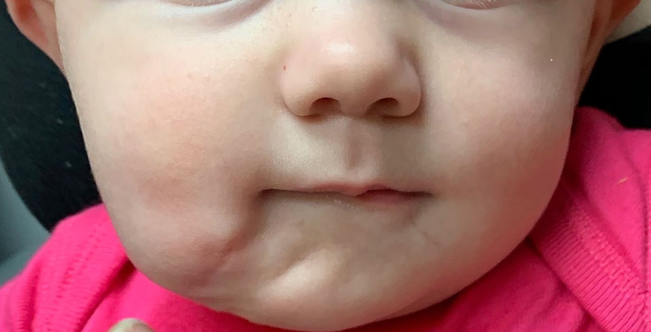
On examination of the newborn, there was a 1–2 cm fullness of the right body of the mandible with intraoral displacement of the tongue to the left (figure 1). A small sinus tract surrounded by vermillion-appearing mucosa was located 1 cm inferior and lateral to the right oral commissure. The sinus tract was approximately 13 mm deep and adjacent to the mandibular mass, with no apparent communication with the oral cavity. The mandibular alveolar ridge was widened several millimetres on the left and she had no ability to depress the right lower lip. The remainder of the head and neck examination was unremarkable. The patient was admitted to the newborn nursery where she exhibited no signs of respiratory distress and demonstrated adequate oral intake prior to discharge.

Image of the face and right mandibular lesion at 5 weeks of age.
At a follow-up appointment at 2 weeks of age, the infant was healthy appearing, feeding well and gaining weight with no oral incompetence. At that time, it was noted that the external component of the mass occasionally developed a raw surface at the skin level that drained clear, serous fluid suspicious for saliva. However, no tests were performed on the fluid. A small accessory tongue appeared to protrude from the opening of the sinus tract and was noted to move in synchronisation with the oral tongue when the infant was feeding.
Investigations
MRI of the face with and without contrast was obtained shortly after birth. This revealed an expansile T1 and T2 hypo-intense lesion to iso-intense lesion arising from the right mandibular body, measuring roughly 2.5×2.1×2.1 cm. The anterior-inferior portion of the lesion, which was likely a soft tissue component of the mass, appeared to track anteriorly and communicate with the subcutaneous surface. The lesion demonstrated several unerupted teeth without evidence of a cystic component or fat to suggest teratoma.
At 2 weeks of age, CT of the mandible without contrast was obtained to further characterise the mass. Similarly, it revealed an ossified and soft tissue mass of the right mandibular body containing teeth (figure 2). There was a soft tissue component along the surface of the bony compartment that extended caudally to the skin surface below the right aspect of the chin. There were no aggressive osseous features or evidence of periosteal reaction.

Three-dimensional CT reconstruction taken at 2 weeks of age.
Treatment
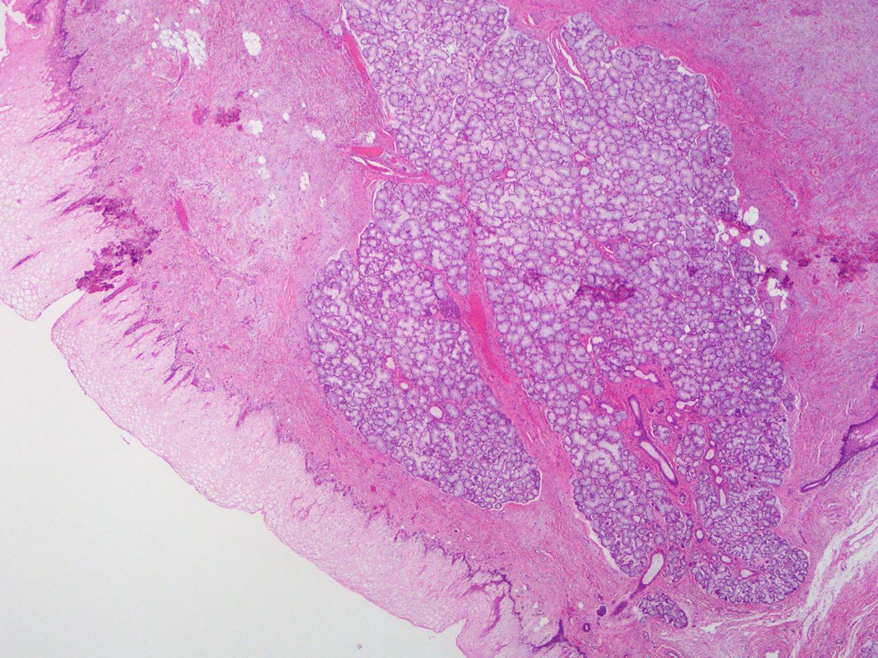
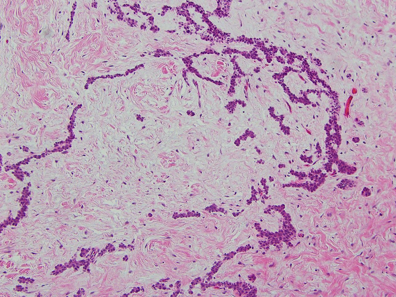
The patient was taken to the operating theatre when she was 6 months old for excision of the duplicated mandible, bony contouring of the mandible and closure of the soft tissue defect with adjacent tissue transfer (figure 3, video 1). Using a combination of blunt and sharp dissection, a plane between the uninvolved soft tissue and the duplicate oral cavity was developed. The mucosal lining and its associated minor salivary glands were resected en bloc and traced to the mandible. The mucosal lining extended onto the mandible and resembled mucosa overlying the alveolar arch. This was peeled off the mandible, and the underlying bone revealed several primary teeth facing toward the duplicate oral cavity. The accessory teeth were extracted and the sockets were drilled down to contour the mandible and remove any remnant dental tissue. Care was taken not to remove any tooth buds that were considered part of her native mandible. Intraoperative nerve monitoring was used to preserve the facial nerve. The remaining soft tissue defect was closed using an advancement flap, which had an area of approximately 8×3 cm. Postoperative pathology revealed a mass consisting of benign squamous mucosa, salivary gland, cortical bone, skeletal muscle and dental pulp with six benign molar teeth (figures 4 and 5).

Operative images of the right mandibular lesion, showing the accessory tongue prior to incision (A), during resection (B) and accessory tooth sockets after resection (C). Image (D) represents the resected mass with associated accessory teeth.

H&E stain at 2× depicting squamous mucosa and salivary gland.

H&E stain at 10× depicting odontogenic epithelial rests.
Outcome and follow-up
Postoperatively, she developed some mild fullness of the right face at the level of the surgical incision for which a repeat CT scan was performed, revealing a fluid collection. The fullness resolved over several months and she did not require further treatment. At 6-month follow-up, the incisions were well healed and the patient was feeding without difficulty but had persistence of the inability to depress the right lower lip, which could represent agenesis of oral depressor muscles or their innervation (figure 6).
The patient’s appearance at 1-month postoperative follow-up.
Discussion
Disopropus, including duplication of stomatodeal structures, is extremely rare, and while phenotypes can range from isolated duplication of cranial or facial structures to a variety of combinations thereof, isolated oral and mandible duplication without associated syndromes have been described only a handful of times.1 2 15 16 This phenomenon was first reported in 1948, where a 2-day-old child was discovered to have a right mandibular mass.17 Similar to our case, the child was noted to have unilateral duplication of the mouth and tongue with a blind pouch and synchronous accessory tongue movement.17 Generally, these craniofacial duplications are diagnosed in-utero or shortly after birth; however, there is one case report from 1978 of an accessory mouth in the temporal region with delayed diagnosis at 22 years old.18 This condition is more commonly reported in females but contributing factors to this demographic have yet to be elucidated and gender differences likely represent a random effect of a rare abnormality.2 10–12 16 19
Chen and Noordhoff proposed a classification system in 1987 for the duplication of stomatodeal structures.20 We have adapted and modified this classification system (table 1).
Classification of stomatodeal structure duplications, adapted and modified from Chen and Noordhoff20
As seen in table 1, association with other anomalies is increasingly more likely with increased classification type. According to this classification scheme, our case represents a type I duplication because our patient had a normal true mouth, a rudimentary duplicated mouth and tongue acting synchronously with the normal tongue and a blind pouch.
There are several theories about the aetiology of this condition, including: duplications of the first branchial arch, anomalies of the notochord including split notochord syndrome, duplication of the mandibular growth centre and abnormal neural crest cell migration.1 21 A more recent study reviewed the available cases of mouth duplication and the authors concluded that duplication of the first branchial arch was the most likely explanation because structures duplicated were all derived from the first branchial arch.22 However, this explanation might not fully explain our case, as other first branchial arch derivatives were normal and there was suggestion of second arch disruption with weakness in muscles supplied by the facial nerve.
Duplication of craniofacial structures typically occurs as part of a syndrome and is often associated with cleft lip and cleft palate, Klippel-Feil syndrome and Pierre Robin sequence.12–14 Cerebral involvement can occur, with duplication of the pituitary gland as the most common manifestation.23 Our patient’s craniofacial duplication is a rare case that presented as an isolated anomaly, with no associated syndromes or abnormalities.
These early duplications can often be identified on prenatal ultrasonography and can be associated with other malformations such as anencephaly, duplications of the central nervous system and neural tube defects.2 A full duplication of all craniofacial structures is often associated with anencephaly and thus incompatible with life. Partial duplications, as in this patient, are unlikely to cause major complications later in life and surgical treatment is a reasonable treatment modality.1 24 If identified in-utero, the priority at time of delivery should be to secure the airway, and an otolaryngologic team should be present at delivery with the necessary equipment. After birth, management depends on the symptomatology of the patient. In our patient, the accessory structures were lateral, they had no communication with the oral cavity and the airway was not compromised.
Imaging should include CT, MRI and possibly angiography to determine structures involved and for perioperative planning purposes.1 3 25 Surgery may be deferred until up to 3 years of age to allow for better differentiation of the teeth, as premature resection may cause inadvertent damage to the unerupted permanent teeth.1
Though this condition is rare, it is important to consider in the differential diagnosis for any unusual mandibular or facial masses seen in infants. There is a risk that these structures could communicate with the oral cavity and involve the airway, placing patients at risk of airway and/or feeding compromise. In a patient with an isolated partial craniofacial duplication and no other anomalies, prognosis is good with definitive surgical resection and reconstruction.
Learning points
Congenital duplication of craniofacial structures is a rare condition that requires close monitoring for a clear understanding of involved structures until the patient reaches the appropriate age for definitive surgical treatment.
Partial craniofacial duplication may exist as an isolated anomaly but is more commonly associated with other craniofacial anomalies and/or syndromes.
CT, MRI or both are helpful prior to surgical intervention for operative planning.
In the case of mandibular involvement, surgical intervention should be deferred until the accessory unerupted teeth can be clearly distinguished on imaging as premature intervention may lead to removal of toothbuds that are a part of the normal mandibular anatomy.
There is a classification by Chen and Noordhoff, adapted and modified for our purposes, that helps elucidate the presentation of these anomalies.20
References
Footnotes
Contributors All listed authors meet all four criteria for authorship as detailed below. AOH: planning, design and drafting of manuscript. DM: planning, design, drafting and editing of manuscript, direct patient care. CC: planning, design of project including direct patient care, photodocumentation and editing of draft. KGP: conception and planning of project including editing of manuscript, acquisition of data and direct patient care.
Funding The authors have not declared a specific grant for this research from any funding agency in the public, commercial or not-for-profit sectors.
Competing interests None declared.
Patient consent for publication Parental/guardian consent obtained.
Provenance and peer review Not commissioned; externally peer reviewed.