Article Text

Abstract
Congenital aplasia of the epiglottis is a rare condition with variable presentation ranging from respiratory distress requiring surgical airway to an asymptomatic finding. Epiglottic aplasia is presumed to be caused by arrest of development of laryngeal structures and is most commonly associated with syndromic conditions, though isolated episodes of aplasia of the epiglottis do exist. In this report, we present a term infant with multiple congenital anomalies who was noted to have a hoarse cry prompting laryngoscopy. This showed complete absence of the epiglottis. Subsequent genetic testing showed mutations in the CPLANE1 gene that is associated with Joubert syndrome. Our patient was able to be discharged home on a thickened formula diet and is eating and gaining weight appropriately. Here, we present a review of the currently available literature of other cases of congenital epiglottic aplasia or hypoplasia discussing the presentation, management and outcomes in these cases.
- ear
- nose and throat/otolaryngology
- otolaryngology / ENT
Statistics from Altmetric.com
Background
Congenital abnormalities of the larynx are rare and are often associated with anomalies of multiple other organ systems and other areas of the upper aerodigestive tract. Primordial functions of the larynx include airway protection with phonation being a later adaptation.1 Abnormalities of the supraglottis are common causes of respiratory distress and feeding difficulties in children with the most common abnormality being laryngomalacia.1 Other anomalies including congenital laryngeal stenoses and atresia are significantly less common. We report the case of a neonate born at term with multiple prenatally diagnosed congenital anomalies. Postnatally, she developed stridor and was noted to have a hoarse cry ultimately leading to the diagnosis of aplasia of the epiglottis.
Case presentation
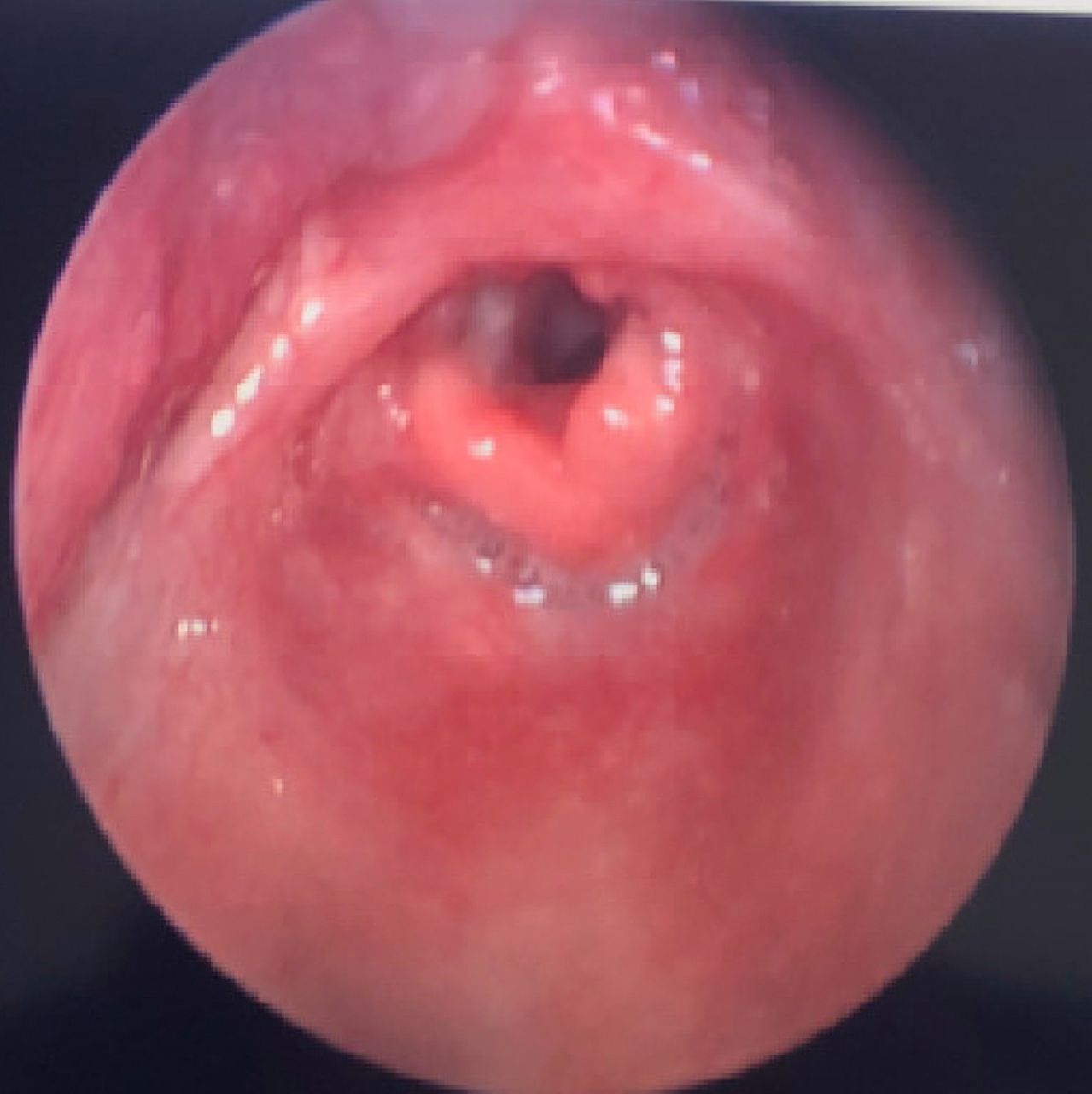
Our patient was born at term to a 34-year-old G2P2002 mother. She had multiple prenatally diagnosed abnormalities including a large atrial septal defect, Dandy-Walker malformation, polydactyly of both feet and the right hand, syndactyly of bilateral feet, dysconjugate gaze, a soft tissue mass on the floor of the mouth and hypotonia. She was originally managed in the newborn nursery but was transferred to higher level of care due to persistent hypoglycaemic. The patient initially did well with oral feeds but began to have difficulty taking in adequate volumes on day of life (DOL) 3 when she began having increased work of breathing and desaturations with feeds. Over the next several days, the primary team and nursing staff noticed intermittent stridor with feeding attempts as well as a hoarse cry for which the otolaryngology team was consulted. Bedside flexible fiberoptic nasolaryngoscopy revealed gross abnormalities including absent epiglottis and hypertrophic arytenoids with diminutive interarytenoid area. The vocal folds demonstrated full adduction and abduction. The patient was subsequently taken to the operating room on DOL 15 for formal microlaryngoscopy and bronchoscopy where the above findings were again confirmed (figure 1). The remainder of the airway examination including the glottis, subglottis, trachea and mainstem bronchi were without abnormalities.
Intraoperative photo from direct laryngoscopy showing absent epiglottis, hypertrophied arytenoids with diminutive interarytenoid Notch.
Differential diagnosis
On initial evaluation of this patient, she was noted to have a hoarse cry but with no audible stridor. The differential diagnosis based on this finding was broad but included unilateral vocal fold paralysis, birth-related trauma causing vagal nerve injury, anatomic abnormality such as laryngeal web or reflux. Bilateral vocal fold paralysis was considered given her stridor and increased work of breathing. In addition, central neurological causes were considered given her known congenital brain abnormalities. Ultimately, diagnosis was confirmed after laryngoscopy showed bilaterally mobile vocal folds with no additional anomalies aside from the absence of the epiglottis.
Treatment
Speech and language pathology was consulted for evalution of stridor with feeds and dysphagia. A modified barium swallow study revealed intratracheal aspiration with thin liquids and laryngeal penetration with nectar thick liquids. She was transitioned to a nectar thickened consistency which she tolerated, though with increased feeding time. She was ultimately discharged home on DOL 18 tolerating full oral feeds with nectar thick consistency.
Outcome and follow-up
At the time of discharge, she was gaining weight appropriately and was tracking along the 66th percentile for weight. At her last follow-up appointment with otolaryngology, she was continuing to gain weight appropriately and was at the 54th percentile for weight. She was continuing to follow her nectar thick consistency diet. Genetic testing was performed given her multiple congenital anomalies. Whole-exome sequencing revealed mutations in the CPLANE1 gene that is associated with autosomal recessive Joubert syndrome.2 This condition is associated with midbrain and hindbrain abnormalities, developmental delay, hypotonia, breathing abnormalities, syndactyly, polydactyly and atypical eye movements.2 A review of the literature reveals only once case of Joubert syndrome associated with epiglottic anomalies. Sung et al report a case of a 2-year-old boy with episodic respiratory distress and stridor. Laryngoscopy revealed absence of the right half of the epiglottis and severe hypoplasia of the left half.3 Of note, our patient’s mother has epilepsy and took oxcarbazepine through the duration of pregnancy. The genetics team believes in utero exposure to this medication may have also contributed to our patient’s congenital anomalies as they cannot all be explained by Joubert syndrome. A review of the literature to date does not reveal any prior publications linking in utero exposure to congenital epiglottic anomalies.
Discussion
Congenital absence of the epiglottis is an exceedingly rare condition and is most commonly described in the setting of syndromic conditions. Less commonly, case reports of isolated epiglottic aplasia have been reported. Manifestations of aplasia or hypoplasia of the epiglottis can vary significantly from some patients being completely asymptomatic, to mild voice changes to early life-threatening dyspnoea requiring tracheostomy tube placement.4 5 A summary of previously published reports of congenital aplasia (table 1) or hypoplasia of the epiglottis (table 2) and overall outcomes of the involved patients are reported below.
Cases of congenital aplasia of the epiglottis
Cases of congenital hypoplasia of the epiglottis
The function of the epiglottis remains controversial. While many believe it plays a role in protecting the airway while swallowing, some studies have shown that it plays a minor role when compared with the true and false vocal cords in airway protection. In addition, case series have shown that adult patients undergoing epiglottectomy for oncological purposes initially developed mild aspiration postoperatively, but this universally resolved after only a few days of speech therapy.6 In our review of cases of congenital absence of the epiglottis, patients exhibit a range of symptom severity from respiratory distress at birth and severe aspiration pneumonia to being asymptomatic into adulthood with epiglottic absence being noted incidentally.
The embryologic development of the larynx is complex. The larynx first appears between days 25 and 28 gestation corresponding to Carnegie stage 10.7 At this point, a thickening of the epithelium forms along the ventral foregut called the respiratory primordium. Around 33 days (Carnegie stage 15), the epiglottic swelling becomes visible in the region of the hypobranchial eminence. The epiglottis is readily identifiable by 41 days gestation and shortly after, demarcates from the base of tongue forming a concave configuration. By the 57th gestational day, the features of the adult larynx are present, but the epiglottis remains unchondrified until the 5th month of gestational age.7 Arrest of development is likely responsible for aplasia and hypoplasia of the epiglottis. Given the fact that other aspects of the larynx are developing synchronously, additional anomalies of the larynx are common in patients with epiglottic anomalies. Thus, we recommend complete airway evaluation to assess for additional anomalies in patients noted to have congenital anomalies of the epiglottis. Interestingly, the hand plate appears at 33 days gestation around the same time that the epiglottis begins to form. This may explain the association between extremity and epiglottic congenital anomalies as seen in our patient and others.7
As detailed in tables 1 and 2, the presentation of patients with congenital epiglottic abnormalities is incredibly variable. While a majority of cases are associated with other congenital anomalies and defined syndromes, there are also additional reports of isolated cases of epiglottic aplasia and hypoplasia, some of which were discovered incidentally in adulthood. Table 3 details cases where infants presented with immediate stridor requiring intubation and ultimately tracheostomy tube placement with variable rates of decannulation with age.8–10 It should be noted in all of these cases that these infants also had additional congenital anomalies which possibly contributed to their initial critical condition. Almost universally when epiglottic anomalies were noted in infancy, the child exhibited feeding difficulties and required either gastrostomy tube placement, nasogastric tube placement or dietary modifications with thickened consistencies as in the case we present. Table 4 details cases where patients required alternative means of enteral access due to feeding intolerance related to their epiglottic anomalies. Feeding difficulty was a much more common manifestation in infants with congenital epiglottic anomalies and was associated with both syndromic and non-syndromic presentations. In contrast, Hong, Kim, Mohamad, Roh and Shahi showed that patients with similar laryngeal anomalies can be relatively asymptomatic and unaffected by their epiglottic anomalies as these patients all presented in adulthood with minor complaints including throat clearing and mild hoarseness.4 5 11–13 These patients all developed compensatory swallowing mechanisms and did not exhibit issues with aspiration.
Cases of congenital epiglottic anomalies where patients required establishment of a surgical airway due to symptom severity
Cases of congenital epiglottic anomalies where patients required alternative means of enteral access due to dysphagia
In this report, we present the case of an infant with likely Joubert syndrome as well as in utero exposure to oxcarbazepine who was noted to have absence of the epiglottis shortly after birth. To our knowledge, only one report exists detailing a congenital epiglottic anomaly in a child with Joubert syndrome and no such reports exist regarding an association between oxcarbazepine and epiglottic anomalies. Our patient’s presentation was relatively mild as she did not develop respiratory distress and was able to tolerate an oral diet with only slight modifications. This is in contrast to the majority of infants diagnosed with epiglottic anomalies at birth who require alternative means of enteral access. At the time of publication, our patient continues to do well with weight gain with no evidence of aspiration and no respiratory difficulties.
Learning points
Congenital epiglottic aplasia and hypoplasia are rare anomalies with presentations that can range from asymptomatic to emergent respiratory distress requiring placement of a surgical airway.
Infants with congenital epiglottic abnormalities do not inherently have respiratory difficulties, nor do they always require surgical intervention to secure a safe airway. Much more commonly, these infants require alternative means of enteral access due to dysphagia and aspiration
The majority of cases of epiglottic aplasia and hypoplasia are associated with syndromic conditions. Patients with epiglottic aplasia should be evaluated for additional congenital anomalies.
Patients with congenital epiglottic anomalies should have a complete swallowing evaluation to monitor for aspiration. Growth should be monitored closely, and alternate forms of enteral access should be obtained when necessary.
References
Footnotes
Contributors LS contributed primary authorship of the original manuscript as well as revisions of further drafts. JD contributed to conceptualisation of the project, production of tables and refinements of drafts. CC contributed to conceptualisation of the project, draft organisation and refinements of manuscript.
Funding The authors have not declared a specific grant for this research from any funding agency in the public, commercial or not-for-profit sectors.
Competing interests None declared.
Patient consent for publication Parental/guardian consent obtained.
Provenance and peer review Not commissioned; externally peer reviewed.